
Brooklyn Physical Therapy News- Evolve NY

Rehabbing Not Resting Painful Tendons
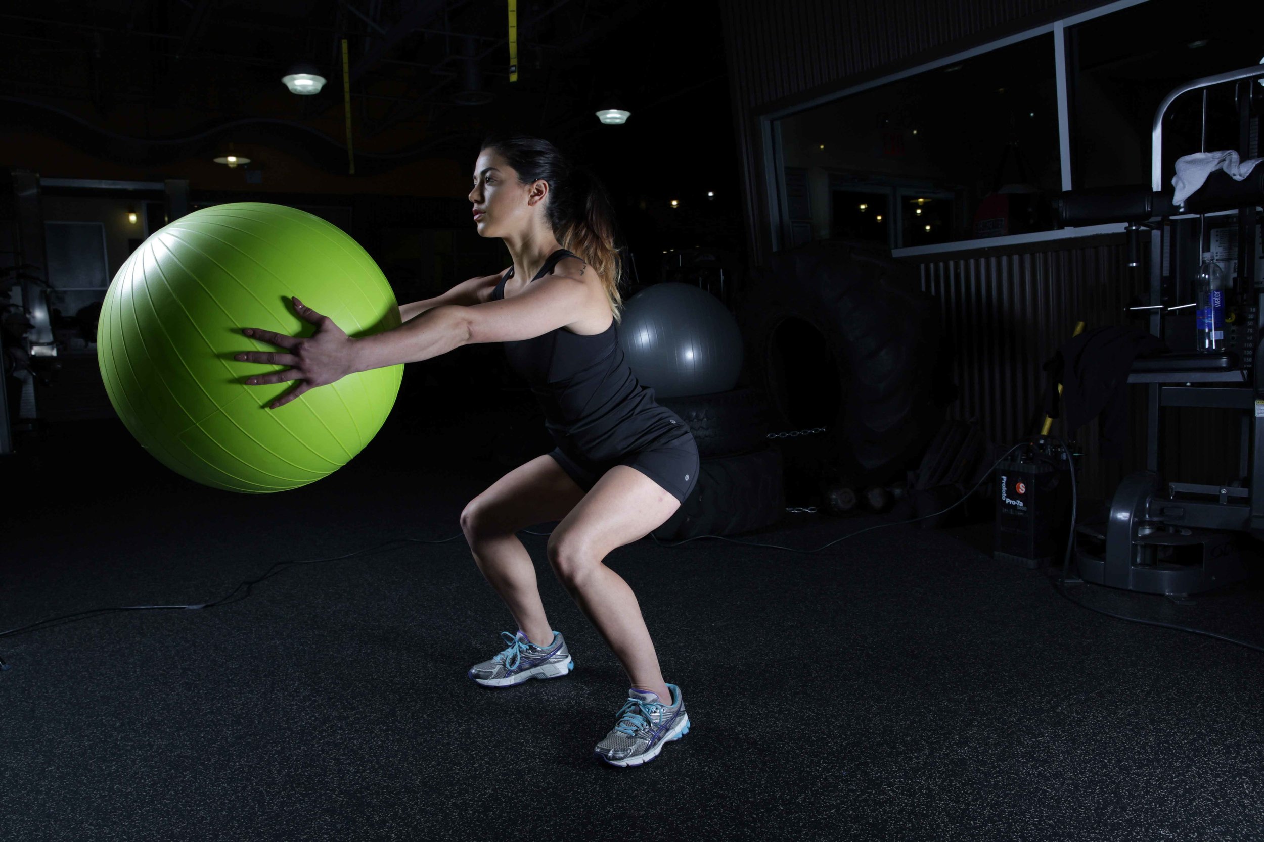
IT’S NOT ALL ABOUT THE MUSCLES: Muscles get most of the attention when it comes to strength and conditioning. We work hard to build those muscles up so that we can lift heavier objects, run faster, jump higher, and look good while doing it. But while our muscles display all of our hard work, our tendons are in the background working just as diligently with little of the glory.
Physical Therapy for Tendonitis
Tackling Tendon Pain Once and For all

Muscles get most of the attention when it comes to strength and conditioning. We work hard to build those muscles up so that we can lift heavier objects, run faster, jump higher, and look good while doing it. But while our muscles display all of our hard work, our tendons are in the background working just as diligently with little of the glory. Tendons are a type of fibrous connective tissue that connect muscles to bones. Far from simply fasteners of muscle to bone, tendons play a pivotal role in the effects that muscles have on the skeleton. First, tendons help to move our bones and amplify the effect of muscle contractions . Second, they play an important role in attenuating and absorbing shock that is transmitted from bone into muscle.
Tendons are strong but they are not designed to stretch or deform easily. They can also become weaker or stronger/stiffer in response to a training stimulus. Depending on the training and the activity, a tendon can become more prone to or more resilient against injury. Tendons are built to withstand a lot of force but repetitive stress which exceeds the tolerance of the tendon can lead to injury. The overloading of the tendon may be caused by ramping up the volume of a new activity too quickly, or due to muscle strength or length impairments, movement pattern inefficiencies and biomechanical errors in and around the involved tendon.
One type of tendon injury that can affect tendons throughout the body is tendonitis. This condition is commonly treated by physical therapists. While other joint conditions can mimic tendonitis, signs and symptoms suggestive of tendonitis include:
Pain over the involved tendon when the muscle is contracted
Pain over the involved tendon when the muscle/tendon is stretched
Swelling or inflammation over the injured tendon
Stiffness when moving the joint near the involved tendon
A feeling of weakness when contracting the involved muscle
Technically any tendon in the body could experience tendonitis, however, certain tendons seem to be more susceptible to this condition than others. Common types of tendonitis include:
Patellar tendonitis (aka jumper’s knee)
Rotator cuff tendonitis
Lateral epicondylitis (aka tennis elbow)
Medial epicondylitis (aka golfer’s elbow)
Achilles tendonitis
Thumb tendonitis (aka De Quervain's tenosynovitis)
Hip flexor tendonitis
Posterior tibialis tendonitis
Hamstring tendonitis
A CASE OF MISTAKEN IDENTITY?
The term “tendonitis” is commonly used to describe pain and irritation of a tendon, but are we using this term incorrectly? It turns out, in many cases, yes. The suffix “itis” denotes the presence of inflammation. In the case of tendonitis, the term suggests the tendon is inflamed, and in the early stages of tendon injury, inflammation is likely present. Except in mild cases of tendonitis which heal quickly, however, the symptoms of a chronic or prolonged case of tendonitis likely have little to do with inflammation and more to do with degeneration that can develop in an injured tendon. This is why it is often more appropriate to refer to a case of persistent tendonitis as a tendinopathy which means “disease or disorder of the tendon.”
Are we splitting hairs here? It may seem that way but understanding what is happening at the level of the injured tissue helps us to select the appropriate treatment interventions. A tendon that is inflamed benefits from treatments aimed at reducing inflammation. A tendon that is degenerating needs a different type of treatment. This is why a tendon that is allowed to rest and treated with ice or anti-inflammatories may recover quickly if caught early. These treatments, however, become less effective over time, and a case of persistent tendinopathy is much more responsive to treatments that improve the stiffness and strength of the tendon rather than simply trying to manage symptoms by reducing inflammation. Similarly, reducing the offending activity can be a good choice early on to allow inflammation to subside and the body’s natural healing process to take over, but prolonged rest may do more harm than good.
Hopefully by now you understand a bit more about the pathophysiology of tendonitis/tendinopathy but you may be wondering how you would know which treatment approach to take. The good news is, you don’t have to figure that out on your own. Physical therapists are experts at musculoskeletal injuries like the one we are describing and are there to tell you which type of treatment or exercise you need. If they believe your tendon injury is in the inflammatory stage they will teach you how to rest and protect the tendon while applying interventions to minimize pain and inflammation. When the tendon has moved beyond the inflammatory stage, your therapist will prescribe treatments and exercises to help increase the stiffness of the tendon so that it becomes more resilient to the repetitive forces of your work tasks and favorite sports. Resting the tendon will not contribute to this process and will predispose you to experience recurrence of these symptoms when you resume your previous level of activities. There is a right way and a wrong way to go about this process of applying graded stress to the tendon and it is a good idea to work with a PT who can help you progress without setbacks.
Tendonitis can seem like a big hurdle to overcome, but with the right treatment plan and the keen eye of a skilled physical therapist, you can get long lasting results that will allow you to return to the activities you love and need to do without worrying that your symptoms will come back as soon as you do. If you are experiencing tendonitis or tendinopathy, let our team of physical therapists at Evolve show you how to recover more quickly and completely.
Click here for more information about physical therapy for tendonitis
About EvolveNY-

Brooklyn's Premier Holistic Physical Therapy Clinics- There’s physical therapy, there’s training, and then there’s EVOLVE. We use the science of biomechanics merged with fitness to help our patients get better and stay better! First we evaluate, then we heal, then we strengthen our clients so they can reach their goals, feel better, and live happier lives. We do so by utilizing a range of core techniques and specialized treatments to reduce pain, improve mobility, enhance physical strength and deal with the underlying issues, not just the pain itself.
Multiple Brooklyn Physical Therapy Locations!
https://EvolveNY.com

Don’t Run Away from Shin Splints
Get Help Healing from Shin Splints for Good- If you are running, jumping and sprinting at high volumes and begin to experience pain along the inside of the shin bone, you may be developing shin splints. While it is tempting to ignore the discomfort and push through your training, shin splints is a condition to be taken seriously and a physical therapist trained in sports and orthopedic injuries can help you recover fully.
Shin Splint Physical Therapy

If you are running, jumping and sprinting at high volumes and begin to experience pain along the inside of the shin bone, you may be developing shin splints. While it is tempting to ignore the discomfort and push through your training, shin splints is a condition to be taken seriously and a physical therapist trained in sports and orthopedic injuries can help you recover fully. Read on to learn more about this condition and why you want a physical therapist in your treatment corner.
MEDIAL TIBIAL STRESS SYNDROME
The medical term for shin splints is medial tibial stress syndrome. This longer name helps explain exactly what is happening when shin splints develop. “Medial tibial” refers to the inner side or medial side of the tibia/lower leg bone which is often referred to as the shin bone. The muscles and tendons involved in shin splints attach along the middle or lower part of the inside of the tibial bone. The term “stress syndrome” denotes that shin splints are an overuse syndrome caused by repeated stress to the tendons and bone in that area.
Shin splints are not the only cause of pain along the inside of the lower leg but should be considered by healthcare providers when a patient presents with complaints of pain along the middle or bottom third of the inside of the tibia. Sharp pain may be felt when their PT or doctor presses along the affected area and a deep ache in that area may be experienced during activity. In the early stages of shin splints the discomfort is present at the beginning of exercise and usually lessens or disappears as an athlete warms up. As it progresses though, symptoms will persist during activity and eventually can become bothersome even at rest.
Unlike a traumatic injury to the tibia like falling and breaking or hitting the bone, medial tibial stress syndrome is considered an overuse injury that usually develops in athletes, military personnel or active persons who run, jump, or sprint at high volumes. Dancers, runners, tennis players and basketball players are some of the athletes at highest risk for developing shin splints. They may notice the onset of these symptoms after increasing the volume or time spent performing these activities.
SHIN SPLINTS ARE MORE THAN A MUSCLE PROBLEM
So what is happening to the lower leg in the case of shin splints? Shin splints are the result of microdamage that accumulates along the attachment point of several lower leg muscles. Repeated contractions of these muscles, such as that which occurs in a runner training for a marathon or a dancer rehearsing for a show, pull on the outer layer of the tibial bone causing microdamage and inflammation of the bone. Without sufficient time for the bone to recover between bouts of activity, this microdamage can accumulate leading to the development of medial tibial stress syndrome or shin splints. Most commonly the posterior tibialis, soleus and flexor digitorum longus muscles are implicated in this condition.
Many athletes will try to push through the discomfort of injuries and conditions like shin splints but we want to help you understand why you should take treating this condition seriously. Unlike other muscle or tendon conditions like a muscle strain or tendonitis, medial tibial stress syndrome is considered to be an early bone stress syndrome. The repetitive pulling of the muscles involved in shin splints along their attachments to the medial tibia bone can begin to cause a stress reaction in the bone. This means that without appropriate modification to activity or technique, shin splints can progress to a bone stress injury or stress fracture.
Bone stress injuries occur in response to repetitive submaximal loading without sufficient recovery time. This repetitive stress to the medial tibia exceeds the bone’s elastic resistance causing structural fatigue and eventually what we know as a stress fracture. Early stress fracture symptoms mimic early shin splint symptoms quite well and it can be extremely difficult to tell how seriously the bone is affected with shin splints. If left untreated, a bone stress injury will worsen and become a significant disruption to your life. Casting or splinting, modified weight bearing and even surgery may be required to promote healing of a stress fracture.
IT’S TIME TO TURN TO YOUR PHYSICAL THERAPIST
Long term healing from shin splints can be tough if you don’t understand what caused the condition to develop in the first place. A physical therapist experienced in orthopedic injuries, however, is a great resource to help you recover from this condition and prevent its return later on. Resting from activity can help reduce the likelihood of shin splints progressing into a full blown bone stress injury but it does nothing to ameliorate the likelihood of symptoms recurring once you return to your previous level of activity.
A physical therapist will ask you about your symptoms, your injury history, your activities and your training schedule to try and identify aspects of your lifestyle or training that may be predisposing you to developing shin splints. They will also perform a physical examination to identify impairments that may be contributing to this condition. For example, the presence of a flattened arch or excessive hip motion during activity can predispose someone to developing shin splints. Limited range of motion in the hip and ankle can also contribute as does the presence of a higher BMI in athletes.
Activity modification, meaning reduction in load bearing activities, is a very important part of allowing the microtrauma involved in shin splints to heal. Stopping all activity, however, is not necessary and may lengthen the time needed to return to full activity. Your PT can guide you through an exercise program to minimize losses in fitness and performance while allowing the bone and tendons to heal. They may utilize modalities like ultrasound or dry needling to promote local healing. While this area is healing your PT will create a treatment plan to improve strength, range or motion, motor control or coordination deficits that may be contributing to the extra stress on your tibia during activity and can recommend a return to activity program that will help you resume activity without reinjury. Dealing with shin splints can be frustrating but with the guidance of a great physical therapist you can feel confident you are taking the best course of action. Call to schedule an evaluation with one of our physical therapists today to learn how you can recover from shin splints for good.
Click here for more information about physical therapy for shin splints
About EvolveNY-
Brooklyn's Premier Holistic Physical Therapy Clinics-
There’s physical therapy, there’s training, and then there’s EVOLVE. We use the science of biomechanics merged with fitness to help our patients get better and stay better! First we evaluate, then we heal, then we strengthen our clients so they can reach their goals, feel better, and live happier lives. We do so by utilizing a range of core techniques and specialized treatments to reduce pain, improve mobility, enhance physical strength and deal with the underlying issues, not just the pain itself.
Multiple Brooklyn Physical Therapy Locations!
https://EvolveNY.com

Physical Therapy for Rotator Cuff
Physical Therapists Tackle Rotator Cuff Tears- Over 2 million people a year in the United States will visit their doctor because of a rotator cuff tear, according to the American Academy of Orthopedic Surgeons. Injuries to the rotator cuff of the shoulder can affect your ability to perform daily tasks like washing your hair, picking up your kids and even sleeping comfortably.
Reviving the Rotator Cuff: Rehab for Rotator Cuff Injuries

Over 2 million people a year in the United States will visit their doctor because of a rotator cuff tear, according to the American Academy of Orthopedic Surgeons. Injuries to the rotator cuff of the shoulder can affect your ability to perform daily tasks like washing your hair, picking up your kids and even sleeping comfortably. The rotator cuff consists of four muscles that play an important role in the function of the shoulder and require different treatment depending on the severity of the tear. In many cases, conservative treatment like physical therapy can be very helpful in managing the symptoms of a rotator cuff tear. If you have a rotator cuff tear or think you may have one, read on to learn more about this injury and how physical therapy can help.
GET TO KNOW THE ROTATOR CUFF
The rotator cuff is a group of four muscles located at the shoulder. The shoulder joint consists of the humerus bone of the upper arm where it meets the glenoid cavity created by the scapula or shoulder blade. The shoulder, or glenohumeral joint as it is anatomically known, is a ball-and-socket joint that allows for a high degree of movement of the arm. The four muscles of the rotator cuff are the supraspinatus, the infraspinatus, the teres minor and the subscapularis. Let’s take a brief look at each muscle and the role that it plays in the shoulder joint.
The subscapularis muscle is located at the top of the shoulder joint. Its muscle belly originates in a hollow area at the top of the scapula or shoulder blade called the supraspinous fossa. Its tendon connects to the upper part of the humerus bone near the shoulder. This muscle aids in lifting the arm up at the side, a movement called “abduction”
Next is the infraspinatus who’s muscle belly lies just below that of the supraspinatus. Its tendon wraps around the outside of the humerus and aids in rotating the arm outward, a movement called “external rotation”.
Just below that muscle, on the scapula, lies the teres minor. This muscle follows the path of the infraspinatus and aids in rotating the arm outward, a movement called “external rotation”.
Finally, the subscapularis muscle actually lies on the underside of the scapula closest to the ribcage. The tendon of this muscle travels through the axilla or underarm to attach near the front of the humerus and aids in rotating the arm inward, a movement called “internal rotation”
As you can see, the muscles of the rotator cuff are essential to many of the movements performed by the shoulder. Not only a mover of the arm, however, the rotator cuff also provides stability to the shoulder joint by helping to keep the head of the humerus centered in the glenoid cavity throughout movement. This is necessary to generate the range of motion and force normally available at the shoulder.
WHEN THE ROTATOR CUFF TEARS
Rotator cuff tears or strains are not uncommon but can present differently depending on the severity of the injury and the location. Any of the four muscles can be injured. Oftentimes rotator cuff tears are the result of progressive wear of the muscle and tendon over time such as with repetitive overhead lifting or work, but it can also result from a single incident such as a fall or accident. A tear can range from minor to severe and may involve the muscle, the tendon or the musculotendinous junction. The presence of pain, swelling and weakness can indicate the severity of the injury. With rotator cuff injuries it is common to experience pain in the shoulder area and pain or weakness when trying to raise the arm to the side, rotate it inward or outward.
Grade I strain (mild): very few muscle fibers are injured. Pain typically occurs the next day but no swelling or bruising is observed. Pain may be felt when the affected muscle is strongly contracted or stretched at its end range.
Grade II strain (moderate): many (but not all) fibers are injured resulting in stiffness, loss of flexibility and loss of strength. Pain is felt both during contraction of the muscle and during stretching. Swelling and bruising over the injured area is common.
Grade III strain (severe): all fibers of the muscle are completely torn or the muscle belly has detached from its tendon. Severe pain is often felt upon injury and heavy swelling and bruising will develop. Range of motion may be either significantly reduced due to pain or excessive because the muscle is no longer limiting it. This muscle will generally be unable to produce any force due to the severe disruption in the fibers.
In the case of a severe strain or tear, a surgical approach may be recommended to restore function to the muscle. Age, functional level and the presence of other surgical risk factors all play a role in the decision to repair a severely torn rotator cuff surgically. In many cases, conservative treatment like physical therapy is recommended to promote healing of injured tissues, reduce pain and inflammation and restore function to the shoulder.
WHAT DOES PHYSICAL THERAPY FOR A ROTATOR CUFF INJURY LOOK LIKE?
During the acute phase of healing which typically refers to the 5-7 days immediately post-injury, the goal of physical therapy will be to protect the site of injury. During this phase, the body will begin to repair the injured tissue but at this point those repairs are very delicate. Any stretching or strong contraction of the injured muscle could re-tear the healing tissue causing the healing process to regress or begin again.Splinting or bracing may be recommended to limit excessive movement or stretching of the muscle and help reduce pain. Application of ice or other modalities like ultrasound or electrical stimulation may be used to reduce pain and promote healing. Passive range of motion performed by your therapist may also be introduced during this time.
As the muscle tear begins to heal your PT may begin to apply very gentle stretching to help maintain tissue flexibility and later on more intensely to restore muscle length. You will begin to perform exercises to restore the motor control and coordination of the muscle initially and the strength and power of the muscle later on. Your physical therapist will help educate you on signs that your muscle is ready to progress to the next stage of rehab and signs that you have overstressed the tissues, such as increased swelling or prolonged pain, and advise you on how to care for it. In later stages of rehabilitation your treatment plan will focus on helping you return to the activities you need to do for life, sports and work.
If you had surgery to repair a severely torn muscle, your physical therapist will help to progress you through your post-surgical protocol with the same goals of managing pain, decreasing swelling, improving flexibility and restoring strength and coordination.
Whether you are an athlete wanting to get back out onto the field or an injured worker trying to get back to your job, working with a skilled physical therapist will help you heal more thoroughly. If you have injured your shoulder, call Evolve PT today to schedule an evaluation with one of our experienced physical therapists.
Click here to find out more information about rotator cuff physical therapy
About EvolveNY-
Brooklyn's Premier Holistic Physical Therapy Clinics- There’s physical therapy, there’s training, and then there’s EVOLVE. We use the science of biomechanics merged with fitness to help our patients get better and stay better!
First we evaluate, then we heal, then we strengthen our clients so they can reach their goals, feel better, and live happier lives. We do so by utilizing a range of core techniques and specialized treatments to reduce pain, improve mobility, enhance physical strength and deal with the underlying issues, not just the pain itself.
Multiple Brooklyn Physical Therapy Locations!
https://EvolveNY.com

Getting to the Bottom of Plantar Fasciitis
Could Your Heel Pain be Plantar Fasciitis? Plantar fasciitis is the most common cause of heel pain in an outpatient setting and can be difficult to treat without the help of a knowledgeable practitioner like a physical therapist.
Could Your Heel Pain be Plantar Fasciitis?

Plantar fasciitis is the most common cause of heel pain in an outpatient setting and can be difficult to treat without the help of a knowledgeable practitioner like a physical therapist. Developing plantar fasciitis can be a bit scary for those experiencing the pain and discomfort of this condition, but after reading this article, not only will you have a better understanding of what is happening when pain in the plantar fascia develops, but also how physical therapy can help you to heal.
GETTING TO KNOW YOUR PLANTAR FASCIA
While most people know about the bones that create the shape of their feet and the muscles that move them, they may be surprised to learn that a third structure, the fascia, plays a pivotal role in how their feet function. The term “plantar” refers to the underside of the foot. The term “fascia” is used to describe a layer of connective tissue made up of closely packed collagen fibers. Fascia is found throughout the body and it envelops muscles, muscle groups, blood vessels, organs and even nerves. Fascia is responsible for holding some structures together while in other places allows for structures to slide and glide along one another. It is flexible but incredibly strong, and can resist tensile forces placed upon it. It also is rich in sensory nerve endings and helps our body know how it is moving and where it is in space.
The plantar fascia originates near the heel on the bottom of the foot and extends all the way to the bones at the ball of the foot. The flexibility and tensile strength of this band of fascia helps to maintain the arch of the foot when you put weight through it. There are three parts to the plantar fascia. The medial component runs along the inside of the foot near the arch. The central component makes up the center of the fascial band and the lateral band runs along the outermost part. There are also some connections between the plantar fascia and the achilles tendon at the base of the calf muscle which also inserts at the heel.
During walking, the plantar fascia stretches some as weight is transferred into the foot. As the heel lifts and weight is transferred over the big toe, the plantar fascia tightens, a process called the windlass mechanism, which helps shorten the foot and possibly conserves energy for forward propulsion. It also acts as a shock absorber for the foot. As you can see, the plantar fascia plays an important role in normal walking mechanics and experiences a great deal of strain and load with each step you take. The role of the plantar fascia in foot mechanics makes it unsurprising that people who spend a lot of time on their feet are more susceptible to developing plantar fasciitis.
PAIN IN THE PLANTAR FASCIA
Now that you know how hard the plantar fascia works to keep you walking, running and jumping, let’s explore what can happen when the plantar fascia becomes injured, a condition known as plantar fasciitis. The suffix “itis” is used to denote the presence of inflammation. Since the condition is called plantar fasciitis, it would be reasonable to think that inflammation of the plantar fascia is the underlying cause of this condition but it turns out inflammation is not the main culprit. Plantar fasciitis is considered to be a mechanical overuse syndrome. Rather than inflammation, the reason for the pain and discomfort experienced with this condition is actually repeated microtrauma or microtears of the fascial fibers. Remember, fascia is highly innervated with sensory receptors which means trauma to those fibers can be quite uncomfortable.
The area of the plantar fascia most susceptible to this microtrauma is near the heel bone which is why your therapist may suspect plantar fasciitis if you present to them with heel pain. Symptoms of plantar fasciitis include pain along the bottom of the heel that is worse in the morning or during the first steps after a prolonged rest. Initially the pain may improve as you warm up but overtime can persist throughout activities. Pain near the heel with prolonged standing or during high impact activities like running, jumping or climbing stairs is also an indicator of plantar fasciitis. It is also often felt while walking barefoot or in shoes with poor support and tenderness may be present along the medial heel.
Pain from plantar fasciitis can range from mild to severe and can impact one’s ability to walk and take part in work, exercise and recreational activities. While we can’t necessarily predict who will develop plantar fasciitis, certain factors can increase one’s risk of developing this condition. Anyone who takes part in prolonged impact activities like distance running or even prolonged standing are more likely to develop this condition than those who spend a lot of time sitting. The presence of certain foot conditions like a high arch (pes cavus) or fallen arch (pes planus) or excessive pronation during gait may also elevate one’s risk. Limitation in ankle or big toe joint dorsiflexion and tightness or weakness in the calf muscle can also be risk factors. Similarly, the presence of a subcalcaneal heel spur, higher BMI (in a nonathletic population) and the presence of Diabetes Mellitus may increase the likelihood of developing plantar fasciitis.
WHAT CAN BE DONE TO ADDRESS PLANTAR FASCIITIS
Like all conditions, getting to the root of the problem is an important step to healing from plantar fasciitis and the earlier you can seek treatment the better. Conservative measures including physical therapy are the first line treatment for plantar fasciitis and can be very effective. Reducing the offending weight bearing activity until symptoms improve is often an important step. Your doctor may recommend the use of NSAIDs or ice to alleviate some pain but your physical therapist will take a multimodal approach to improving your symptoms, allowing the plantar fascia to heal and reducing the likelihood of symptoms recurring. Physical therapy for plantar fasciitis often includes:
Stretching
Dry needling
Strengthening
Night splints or orthotics
Optimizing lower extremity biomechanics during activities like running, jumping and walking
Recommendations for footwear
Manual therapy to the plantar fascia, foot, and ankle
Other modalities to promote tissue healing
More invasive treatments like corticosteroid injections, platelet rich plasma (PRP) injections, and surgical interventions are saved as a last resort for cases that do not respond to conservative treatment. If you are experiencing heel pain or have been diagnosed with plantar fasciitis, you will want to start your recovery off on the right foot. Call Evolve Physical Therapy to schedule an evaluation with one of our experienced physical therapists today.
Click here for more information about physical therapy for plantar fasciitis
About EvolveNY-
Brooklyn's Premier Holistic Physical Therapy Clinics- There’s physical therapy, there’s training, and then there’s EVOLVE. We use the science of biomechanics merged with fitness to help our patients get better and stay better!
First we evaluate, then we heal, then we strengthen our clients so they can reach their goals, feel better, and live happier lives. We do so by utilizing a range of core techniques and specialized treatments to reduce pain, improve mobility, enhance physical strength and deal with the underlying issues, not just the pain itself.
Multiple Brooklyn Physical Therapy Locations!
https://EvolveNY.com

Physical Therapy for Heel Pain
Get Help for Painful Heels! As many as 1 in 10 people will experience pain in their heel or heels at some point in their life. That is to say, heel pain is fairly common but that doesn’t mean you will welcome its presence. In fact, heel pain can range from mildly annoying to quite painful and given that many of us walk as our primary form of mobility, it can be quite disruptive to our lives.
Stop Tip-Toeing Around Heel Pain

Get Help for Painful Heels!
As many as 1 in 10 people will experience pain in their heel or heels at some point in their life. That is to say, heel pain is fairly common but that doesn’t mean you will welcome its presence. In fact, heel pain can range from mildly annoying to quite painful and given that many of us walk as our primary form of mobility, it can be quite disruptive to our lives. If you are dealing with heel pain you are probably wondering what may be the cause. Let’s look a little deeper at the heel itself and discuss some of the common causes of heel pain treated by physical therapists.
ANATOMY OF THE HEEL
The heel refers to the back part of the foot. From a bony standpoint it is composed of the calcaneus bone. The calcaneus sits below the talus, the bone that articulates with the two lower leg bones to form the ankle joint. In front of the calcaneus are the tarsal bones which then connect to the long bones in our foot. The calcaneus is a sturdy bone that supports the weight of our leg and body and plays an important role in walking or gait. It acts as a short lever for our calf muscles to point the foot and bend the knee during walking, running and jumping activities.
The calcaneus provides an insertion point for several muscles and ligaments as it is the largest and broadest of all the foot bones. As mentioned above, the calf muscles (gastrocnemius and soleus) insert along the posterior aspect of the calcaneus via the achilles tendon. Other muscles that attach there are the abductor hallucis (flexes and moves the big toe outward), flexor digitorum brevis (flexes the outer four toes), quadratus plantae (supports the arch and helps flex the toes), abductor digiti minimi (moves the little toe outward) and extensor digitorum and hallucis brevis (extend the toes upward).
Several ligaments that stabilize the foot and ankle attach to the calcaneus on either side and from beneath the foot. The plantar fascia, a thick fibrous band of tissue that runs longitudinally along the bottom of the foot from the base of the heel up toward the toes is also an important structure when it comes to heel pain. It plays an important role in the biomechanics of the foot during walking and standing and in shock absorption. As you can see, there are quite a few places where heel pain can develop.
AM I AT RISK FOR HEEL PAIN?
While heel pain is a fairly common complaint many people will never experience this condition. Certain factors and activities, though, can increase one’s risk for developing heel pain. Lifestyle factors like participating in activities that involve a lot of running or jumping may increase the chance of heel pain or discomfort developing. Similarly, spending a lot of time on your feet, especially on concrete floors or while wearing unsupportive or uncushioned footwear may increase the risk. Finally, intrinsic risk factors like being overweight or having foot arthritis or flat feet may also increase the risk for heel pain. Having any one or several of these factors does not guarantee you will develop heel pain but identifying the risk factors can help provide targets for treatment or prevention.
WHAT MIGHT BE CAUSING YOUR HEEL PAIN?
While there are many potential sources of heel pain, here are some of the more common causes:
Heel spurs: Heel spurs are bone spurs or osteophytes that form on the calcaneal (heel) bone of the foot. These areas of excess bone can cause pain, inflammation, tenderness and joint stiffness in the area.
Sever’s Disease: The most common cause of heel pain in growing children, Sever’s Disease occurs when the growth plate at the back of the heel becomes inflamed and painful.
Plantar fasciitis: When inflamed or irritated pain can be felt in the bottom of the heel, along the arch or at the ball of the foot.
Achilles tendinitis: Achilles tendinitis is one of the most common causes of heel pain felt along the back of the heel. Injury or irritation at the attachment site or distal end of the achilles tendon which connects the calf muscle to the calcaneus is a hallmark sign.
Tarsal Tunnel Syndrome: Entrapment of the tibial nerve where it runs beneath a band of ligaments along the inner ankle can cause pain, numbness, tingling or burning along the sole of the foot, including the heel.
Stress fracture: Calcaneal stress fractures can occur in persons participating in high volumes of weight bearing activities like running or marching.
IDENTIFYING THE CAUSE OF HEEL PAIN
Identifying the cause of heel pain is helpful in developing an effective treatment plan. There are several tools that physical therapists and other medical professionals use to pinpoint the possible causes of heel pain. Some of these tools include:
Subjective interview: Don’t underestimate the importance of describing your symptoms to your healthcare provider. Providing information like where the pain is located, when it started, what provokes and relieves the pain and the characteristics of the pain can be very helpful to healthcare professionals in developing a list of possible causes.
Movement analysis: Watching the way your foot, ankle, knee and hip move while you stand, walk, jump and run can give clues as to what may be provoking your pain. Problems with form and biomechanics during repetitive movements can lead to pain or discomfort in areas that are experiencing higher levels of stress or strain than normal.
Palpation: Gently palpating or pressing along the heel can help your provider locate the exact location of your pain. By identifying the location they can more easily identify the anatomical structures that may be involved in your symptoms.
Imaging: When necessary, imaging studies such as X-rays, MRIs or CT scans may be warranted to look for structural issues and injuries like bone spurs, fractures, ligament tears, tendon inflammation and so forth. It is important to know that while imaging can sometimes be very helpful in locating the source of your pain, other times imaging may reveal what we would consider to be “abnormal” findings that don’t actually cause any symptoms at all. They may be a red herring, so to speak.
While living with heel pain can be quite frustrating, you don’t have to figure it out on your own. Our physical therapists can help you identify the underlying cause of your pain and develop a plan to reduce your discomfort and minimize the risk of recurrence in the future.
Click here for more information about physical therapy for heel pain
About EvolveNY-
Brooklyn's Premier Holistic Physical Therapy Clinics- There’s physical therapy, there’s training, and then there’s EVOLVE. We use the science of biomechanics merged with fitness to help our patients get better and stay better!
First we evaluate, then we heal, then we strengthen our clients so they can reach their goals, feel better, and live happier lives. We do so by utilizing a range of core techniques and specialized treatments to reduce pain, improve mobility, enhance physical strength and deal with the underlying issues, not just the pain itself.
Multiple Brooklyn Physical Therapy Locations!
https://EvolveNY.com

Physical Therapy for Heel Spurs
Physical Therapy for Heel Spurs- If you have been dealing with pain, inflammation or warmth over your heel, you could have a condition called a heel spur. This condition affects up to 1 in 10 adults though many individuals may not even realize they have a heel spur.. The likelihood of finding them on imaging increases with age. For those experiencing discomfort due to the presence of a heel spur, let’s learn more about the condition and how PT can help.
Save the Heel Spurs for Cowboys!
Get Help for Painful Heel Spurs!

If you have been dealing with pain, inflammation or warmth over your heel, you could have a condition called a heel spur. This condition affects up to 1 in 10 adults though many individuals may not even realize they have a heel spur.. The likelihood of finding them on imaging increases with age. For those experiencing discomfort due to the presence of a heel spur, let’s learn more about the condition and how PT can help.
TWO TYPES OF HEEL SPURS
Heel spurs are a type of bone spur named for their location along the calcaneus or heel bone.
They can develop in two different locations along the calcaneal bone. When a bone spur forms along the bottom of the heel on the sole of the foot, it is called bone spur syndrome. Alternatively, a bone spur that forms on the back of the calcaneus where the achilles tendon inserts is called insertional achilles tendonitis. In some cases a bony protuberance can be felt under the skin in these areas.
The medical term for bone spur is osteophyte. The prefix “osteo” means related to bones while the suffix “phyte” refers to pathologic outgrowth. Taken together, the term osteophyte refers to an outgrowth of bone. The body most commonly develops bone spurs in an attempt to repair or address an injury. It does so by producing excess bone and calcium where there has been trauma to the joint, repeated tendonitis, breakdown of the cartilage or repeated overuse of a joint. Does anyone else in your family have bone spurs? Research suggests it may also have a genetic cause
Bone spurs are most common in adults over the age of sixty and usually grow in areas where osteoarthritis–breakdown of the cartilage from wear and tear–has formed in a joint. Bone spurs can grow from any bone but are most common in the foot (especially the heel, big toe and ankle), the hands and fingers, hips, neck and spine, knees and shoulders. In the heel, repeated stretching of the plantar fascia or tearing of the membrane that covers the heel bone can lead to heel spur formation. Runners that run on hard surfaces, those wearing improper or unsupportive footwear when on their feet for long periods and those who are overweight or obese are also at a higher risk for developing heel spurs.
NOT ALL HEEL SPURS CAUSE SYMPTOMS
Oftentimes the presence of a heel spur is an incidental finding when looking for something else. It is important to rule out other causes for foot or heel pain as the presence of a heel spur is not necessarily associated with having symptoms. In the case of heel spur syndrome it is important to differentiate between a case of plantar fasciitis and a symptomatic heel spur. So while some heel spurs are asymptomatic, bone spurs located in an area that can irritate tissue or impede range of motion can cause symptoms. If you are experiencing any of the following symptoms, you may have a heel spur:
Sharp pain or intense ache in the heel that is often worse first thing in the morning or after a longer period of rest
Inflammation or swelling over the heel
Tenderness when touching or applying pressure to the area
Warmth over the area
A palpable bony protrusion in the heel
CONSERVATIVE MANAGEMENT CAN HELP MANAGE HEEL SPURS
Conservative, non-surgical management like physical therapy results in symptom improvement in the majority of patients. By making some of these changes you can help improve the long-term success of your rehabilitation program:
Avoid long periods of time on your feet. Take breaks regularly, especially in the beginning of your treatment when you are more symptomatic
Wear well-fitting shoes with proper arch support when you will be standing or walking for extended periods
Wear slippers or cushioned shoes for walking on tile or hardwood floors at home
Run or walk on softer surfaces outdoors like grass rather than only on hard surfaces like concrete or asphalt.
Heel pads or shoe inserts may be beneficial to help distribute pressure more evenly across the heel
Physical therapy can be a great tool for managing symptomatic heel spurs. While it is true that physical therapy will not eliminate the heel spur, it can help reduce the irritation and inflammation around the spur that is causing your symptoms. As mentioned earlier, many people have heel spurs without any symptoms and we’d like you to be one of those people. Physical therapy aims to reduce inflammation, improve foot and ankle motion, address muscle and soft tissue impairments and improve the quality of movement to minimize heel spur symptoms.
Let's take a look at some exercises that can help manage the symptoms of a heel spur that your PT may prescribe. Please note, it is important to check with your doctor before starting a new exercise plan. Also, these exercises are meant to be only examples of what you may encounter while working with your physical therapist. Your PT will provide you with a customized physical therapy exercise program to treat your heel pain.
Calf stretching: Stand with your hands against the wall in a lunge position. With the back foot pointed straight ahead and knee straight, lunge forward over the front foot until you feel a moderate stretch in the back calf muscle. Hold for 20-30s and perform 2-3 repetitions to help stretch the calf muscle.
Big toe stretch: While seated, grasp your big toe on the symptomatic side and gently pull it back toward you until a gentle stretch is felt along the bottom of your foot. Hold this for 20-30s and perform 2-3 repetitions at a time to help mobilize the big toe and stretch the plantar fascia.
Massage: If your heel spur is on the sole of the heel, massaging the plantar fascia and bottom of the foot may help. Place a massage ball or frozen water bottle under your foot and roll your foot back and forth across the ball or bottle spending extra time in spots that feel tender.
Arch strengthening: Start by sitting with your feet flat on the floor. Place a pen under the arch of your affected foot. Without raising the foot off the ground, use your intrinsic foot muscles to try and lift the arch up and off the pen, holding for a few seconds at the top before lowering back down. Perform 15-20 reps for 1-2 sets to help re-educate and strengthen the muscles that support the arch. Progress to standing when you are ready for a greater challenge.
While in some cases surgery may be necessary to remove heel spurs, a physical therapist at Evolve can help you to reduce pain and swelling, improve your joint mobility and optimize your movement patterns. Give us a call today to schedule an evaluation and learn more about how we can help you manage the symptoms of a heel spur. Call: 1-718-258-3300
Click here for more information about physical therapy for heel spurs
About EvolveNY-
Brooklyn's Premier Physical Therapy Clinics-
There’s physical therapy, there’s training, and then there’s EVOLVE. We use the science of biomechanics merged with fitness to help our patients get better and stay better!
First we evaluate, then we heal, then we strengthen our clients so they can reach their goals, feel better, and live happier lives. We do so by utilizing a range of core techniques and specialized treatments to reduce pain, improve mobility, enhance physical strength and deal with the underlying issues, not just the pain itself.
Multiple Brooklyn Physical Therapy Locations!
https://EvolveNY.com

Breaking Through Bad Bursitis
Bursitis 101: Bursitis is a condition many are unfamiliar with until they begin to experience the telltale signs. Because bursae are located throughout the body, bursitis is not relegated to only one limb or joint but can occur in many different places. When pain is occurring near or around a joint, bursitis is one condition that should be included in the differential.
Physical Therapy for Bursitis
Bursitis 101

Bursitis is a condition many are unfamiliar with until they begin to experience the telltale signs. Because bursae are located throughout the body, bursitis is not relegated to only one limb or joint but can occur in many different places. When pain is occurring near or around a joint, bursitis is one condition that should be included in the differential. Let’s take a deeper look at bursitis and how this condition can present in different parts of the body.
SO MANY BURSAE…
Bursae (plural of bursa) are small fluid-filled sacs found near joints. Healthy bursae are very thin. They consist of a synovial outer membrane filled with slippery synovial fluid. These slippery sacs help the tendons, ligaments, muscles and skin glide smoothly over the bony surfaces of the joints. Though they contain only a small amount of fluid, they provide some cushioning to the area. These bursae play such an important role that there are actually more than 150 of them in the body.
Bursae are located throughout the body. The major bursae commonly affected by bursitis are located near the joints of the shoulders, elbow, hips and knees, though technically bursae anywhere can become inflamed. Because of the bursae’s location beneath and between tendons, ligaments, muscles and skin, repeated movement over a bursa or prolonged pressure or positioning can result in inflammation of the bursa known as bursitis. As inflammation sets in, more synovial fluid is produced causing the normally thin sac to swell and take up even more space than they usually do. When this happens, many of the common symptoms of bursitis can develop.
Common symptoms of bursitis include pain and tenderness in the area of the inflamed bursa. It may be difficult to lie on the area of the body with bursitis due to the discomfort it causes. Joint mobility may be limited in the area around the inflamed bursa. If the bursa is located just below the surface of the skin you may even be able to visualize some redness and swelling in the area. Some people will experience repeated bouts of bursitis with resolution of symptoms in between.
AM I AT RISK FOR DEVELOPING BURSITIS?
Both lifestyle habits and the presence of certain medical conditions can put one at risk for developing bursitis. Let’s take a look at how some activities or lifestyle habits may affect certain bursa:
Olecranon bursitis: The olecranon is the bony protuberance at the back of your elbow. Spend too much time leaning on your elbow on the desk while you work and you could inflame the bursa that lies just underneath the skin
Trochanteric bursitis: The greater trochanter is located on the lateral side of the thigh near the hip. Running and frequent stair climbing may irritate the bursa at the greater trochanter where several muscles attach that move the hip and leg.
Subacromial bursitis: The acromion is the front portion of your shoulder blade that protrudes across the top of your shoulder. Beneath the acromion and above the tendons that connect there lies the subacromial bursa that can become irritated and inflamed with repeated overhead motions such as throwing or reaching to stock high shelves.
Infrapatellar bursitis: One of the bursa located beneath the patellar tendon of the knee where it attaches on your lower leg, the infrapatellar bursa may also experience bursitis. Also known as “clergyman’s” knee, this bursa can become inflamed as a result of direct trauma to the patella or kneecap, from frequent kneeling and even overuse.
Being diagnosed with certain medical conditions that can affect the joints may also put you at risk for developing bursitis. These conditions include osteoarthritis, diabetes, rheumatoid arthritis and gout. In some cases it may be difficult to tell whether pain is coming from the joint itself or the nearby bursa.
Finally, septic bursitis is a less common but more serious cause of bursitis. In this case the bursa becomes infected by bacteria that has entered through a cut, scrape or abrasion on the skin. Rapid identification of this condition is important to allow for initiation of proper treatment.
WHAT ARE MY TREATMENT OPTIONS
Treatment of bursitis is typically conservative in nature. There are several treatment options, some which require the help of a physician. Let’s explore some common treatment options for bursitis.
Anti-inflammatories: Your doctor may recommend the use of anti-inflammatory medications to help calm the inflammation and reduce your pain. This may be prescribed in addition to other strategies like activity modification or the use of ice.
Corticosteroid injections: If you visit your doctor’s office for bursitis they may recommend that you undergo a corticosteroid and lidocaine injection into the affected bursa. This combination of medication can quickly reduce the inflammation and local pain.
Fluid aspiration: In some cases, when other options have failed, your physician may elect to aspirate the fluid that has built up in the bursa.
Physical therapy: Physical therapy is an important tool for treating bursitis. While the use of anti-inflammatories and corticosteroid injections can provide you with relief of your symptoms, it does not address the underlying cause of the bursitis thus placing you at elevated risk for developing future episodes.
With physical therapy, the goal is not only to address your immediate symptoms of pain and discomfort but to get to the root cause of the irritation on that bursa. Oftentimes optimizing flexibility and tissue mobility, muscle strength and coordination, motor control and balance along with some modifications to training or technique can provide long-term relief of symptoms.
Decreasing the pressure and irritation on the bursa while continuing to participate in the activities you need and love to do is physical therapy’s goal. So while you may be feeling discouraged from a bout or several bouts of bursitis, our team of skilled physical therapists at Evolve is here to help you onto the road to long-term symptom relief.
Click here for more information about physical therapy for Bursitis
About EvolveNY-
Brooklyn's Premier Holistic Physical Therapy Clinics- There’s physical therapy, there’s training, and then there’s EVOLVE. We use the science of biomechanics merged with fitness to help our patients get better and stay better! First we evaluate, then we heal, then we strengthen our clients so they can reach their goals, feel better, and live happier lives. We do so by utilizing a range of core techniques and specialized treatments to reduce pain, improve mobility, enhance physical strength and deal with the underlying issues, not just the pain itself.
Multiple Brooklyn Physical Therapy Locations!
https://EvolveNY.com

Physical Therapy for Torn Meniscus
MEET THE MENISCUS : The menisci (plural for meniscus) are small but important structures found in the knee. So important, in fact, that you have not one but two menisci on each side. The knee, anatomically known as the tibio-femoral joint, consists of the articulation between the end of the femur, or long thigh bone, and the flattened top portion of the lower leg or tibia bone.
Physical therapy for Torn Meniscus
An Updated Approach to Treating Meniscus Tears
A 15 year old soccer player is running down the field toward the goal. The ball comes their way and they plant their left foot and turn to kick the ball with their right. The ball sails into the goal but the player feels a sharp pain in their left knee as they jump up to celebrate.
What happened? Well, they may have just torn their meniscus.
MEET THE MENISCUS
The menisci (plural for meniscus) are small but important structures found in the knee. So important, in fact, that you have not one but two menisci on each side. The knee, anatomically known as the tibio-femoral joint, consists of the articulation between the end of the femur, or long thigh bone, and the flattened top portion of the lower leg or tibia bone. As you probably know, the job of the knee joint is to bend and straighten while you walk, run, jump and squat. There are quite a few structures within the knee that provide it strength and stability. One–or rather two–of those structures are the menisci.
The menisci are wedged, c-shaped discs anchored onto the flattened end of the tibia bone. There is one on the medial (inner) side and one on the lateral (outer) side and they curve toward one another. Their job is both to act as shock absorbers/cushions in the knee and to help stabilize the knee joint. Because of their location within the joint, these little cushions are subject to the stress and strain of all of the activities we do throughout our life while on our feet.
HOW DO MENISCI GET INJURED?
If you think about all of the running, jumping, squatting, and stair climbing you’ve done throughout your life so far, you can imagine that the knees take on a lot of stress. Meniscus injuries, like any injury, occur when the stress or strain on the tissues of the meniscus becomes too great. When this happens, the meniscus can tear. Tears can be categorized as acute/traumatic tears which usually occur when leg is twisting while the foot is planted or as degenerative tears which occur in response to wear and tear over time. In either case, meniscus tears can be uncomfortable and painful and interfere with a person’s ability to walk and be active.
HOW DO I KNOW IF I’VE INJURED MY MENISCUS?
Meniscus injuries are definitively diagnosed using imaging technology like MRI or CT scan to visualize the tear. Clinically, however, we look for certain signs and symptoms such as:
An audible or felt pop
Pain in the knee joint on the inside (medial), outside (lateral), or back of the knee
Swelling
Catching or locking of the knee joint
Pain with walking
Inability to fully bend or straighten the knee
Pain when twisting the knee
Feelings of the knee giving out
These are signs and symptoms of a symptomatic meniscus tear but guess what? Not all meniscus tears are symptomatic. In fact, imaging studies have found incidental and asymptomatic meniscus tears in up to 5% of young people and up to 67% of older adults. In fact, the presence of a meniscus tear on imaging is not predictive of knee symptoms in middle age or older adults. It is associated with the presence of osteoarthritis in older adults, but not directly linked to symptoms. This is more evidence that as physical therapists, we treat symptoms, not imaging.
WHEN DO I NEED TO SEEK TREATMENT?
If you are experiencing knee pain or stiffness, locking or catching, instability or swelling it is time to have your knee examined. Physical therapists assess the knee through clinical tests like the Apley’s or McMurray’s tests and through palpation and examination of the knee. In the case of an acute or traumatic tear, the mechanism of injury combined with the symptom report may point your PT in the direction of suspecting a meniscus tear but they will also examine other structures in the knee. Due to the mechanism of injury with a meniscus tear, other structures in the knee such as the anterior cruciate ligament (ACL) or medial collateral ligament (MCL) may be injured at the same time. If your PT suspects a torn meniscus they will likely refer you to an orthopedist for further assessment.
PEACE & LOVE FOR MENISCUS TEARS
The acronym R.I.C.E used to be the standard of early treatment for an acute injury like a meniscus tear.
R: Rest
I: Ice
C: Compression
E: Elevation
Current practice guidelines continue to evolve, and many therapists now preach P.E.A.C.E. & L.O.V.E., baby. This creative acronym outlines a new, more active approach to addressing soft tissue injuries like a meniscus tear and you may encounter this approach you work with your physical therapist. Let’s take a deeper look at it. (*Note, this does not apply directly to post-surgical cases and as every case is different, your therapist or physician may still recommend ice and anti-inflammatories to manage pain and swelling)
(P)rotection: Protecting the injury in the first week or so is important to minimize disruption of early healing tissues which are still vulnerable. This includes avoiding activities that increase pain and swelling and sometimes modifying weight bearing with crutches or a walker.
(E)levate: elevate the limb above the heart as often as possible to allow swelling to drain from the area. You can achieve this by lying down and raising your leg on a stack of pillows.
(A)void taking anti-inflammatories: Unless recommended by your physician for other reasons, it may be beneficial to avoid interfering with the inflammatory process which is actually a necessary part of the healing process as it helps clear away unwanted debris and usher in helpful cells. This can apply to ice as well.
(C)ompression: apply an elastic band or taping around the knee to minimize swelling
(E)ducation: Your PT will educate you on how to protect the knee during the early healing phase and how to support your body in it’s natural healing process
AND
(L)oad: Gradually loading the tissues is important after the first week. The presence or absence of pain and inflammation will be your guide.
(O)ptimism: focus on what you can do and remain optimistic about your body’s power for healing
(V)ascularization: choosing pain-free cardiovascular training will increase blood flow to healing tissues
(E)xercise: Graded exercises should be used to help restore normal strength, balance, flexibility and proprioception in accordance with your stage or healing
While this acronym may seem a bit complicated, you won’t have to go through it alone. If you are dealing with a torn meniscus or think you may be, schedule an evaluation with one of our knowledgeable physical therapists and we will help you get on the road to recovery.
Click here to find out more information about physical therapy for a torn meniscus
About EvolveNY-
Brooklyn's Premier Holistic Physical Therapy Clinics- There’s physical therapy, there’s training, and then there’s EVOLVE. We use the science of biomechanics merged with fitness to help our patients get better and stay better!
First we evaluate, then we heal, then we strengthen our clients so they can reach their goals, feel better, and live happier lives. We do so by utilizing a range of core techniques and specialized treatments to reduce pain, improve mobility, enhance physical strength and deal with the underlying issues, not just the pain itself.
Multiple Brooklyn Physical Therapy Locations!
https://EvolveNY.com

Physical Therapy for Spondylosis
Spondylosis: Wearing out its Welcome- Under the best of circumstances getting older means you have created a lifetime of good memories. Hopefully your body has carried you through lessons and adventures you can pass along to the next generation. With aging, however, our physical bodies undergo changes that can lead to sensations of pain, stiffness and even weakness.
Spondylosis: Wearing out its Welcome

Under the best of circumstances getting older means you have created a lifetime of good memories. Hopefully your body has carried you through lessons and adventures you can pass along to the next generation. With aging, however, our physical bodies undergo changes that can lead to sensations of pain, stiffness and even weakness. While this may seem like an inevitability, there is often a lot that can be done to alleviate these symptoms so that you can continue living your best life. As physical therapists, healthy aging is something we are passionate about and this article describes how physical therapists can address symptoms of spondylosis, an umbrella term for a number of degenerative changes that can occur in the spine.
WHAT WE MEAN WHEN WE SAY “SPONDYLOSIS”
Spondy- meaning “spine” and -losis meaning “condition or state of” gives us a generic term meaning condition of the spine. In the medical world, spondylosis is an umbrella term used to describe age-related changes that can occur in the spine, sometimes referred to as “wear and tear.” These degenerative changes can affect some or many of the different structures found in the spine such as the intervertebral discs, the ligaments in the spine and even the bony structures themselves.
Intervertebral discs become more dry and may crack and lose height, ligaments can thicken and cartilage may become worn down. Spinal stenosis in which the spinal canal or vertebral foramina are narrowed causing pressure or irritation of the spinal cord and spinal nerves may develop as a result of bone spur formation. Discs may be more likely to herniate causing pain and discomfort in the back and potentially into more distant areas of the upper and lower extremities. Finally movement may not be as smooth secondary to loss of cartilage.
The only way to truly identify these age-related changes is through imaging such as radiographs, CT scans or MRIs. It is interesting to note, however, that changes such as these can be observed in imaging of pain-free adults as well as adults experiencing pain. This is to say that the presence of spondylosis does not mean that you will experience pain or discomfort. The presence of these degenerative changes combined with other factors such as imbalances in muscle strength and length, changes in joint mobility and flexibility, patterns of movement and habits of movement may lead to the development of these symptoms, however. These factors are often the targets of physical therapy intervention to address spondylosis-related symptoms.
PHYSICAL THERAPY CAN IMPROVE SYMPTOMS OF SPONDYLOSIS
Physical therapists are skilled movement practitioners. They aim to address symptoms like pain, stiffness or weakness so that you can continue doing the activities that you love and need to do. This approach applies to spondylosis as well. If you are considering physical therapy to address some of your spondylosis symptoms, you may be wondering how physical therapists approach this condition. While an individualized approach is always best, let’s take a look at how physical therapists commonly address spondylosis:
Reduce pain, inflammation and irritation: While completely eliminating your symptoms may not be realistic in all cases, providing as much relief as possible is an important goal of physical therapy for spondylosis. In many cases, repeated irritation of involved structures results in symptoms of spondylosis. For example, in the case of a herniated intervertebral disc, irritation of the spinal nerves near the disc can cause localized and radiating pain, numbness or tingling. Similarly, the presence of a bone spur may irritate the spinal cord when the spine is moved or held in certain positions.
Reduction of pain, inflammation and irritation is a multi-step process. One step involves addressing local symptoms of pain, muscle stiffness or spasm and inflammation through modalities like ice, heat, electrical stimulation, soft tissue mobilization or dry needling. The other steps involve identifying and addressing patterns of movement or impairments in muscle strength, motor control, coordination, and flexibility, for example, that may contribute to increased irritation or stress across the area affected by spondylosis. Addressing and optimizing these components can help decrease the likelihood that the spondylotic changes in the spine will result in unwanted symptoms.
Movement is key
While there are certainly times when rest is necessary for recovery such as after an acute injury or surgery, in many other cases too much rest can be counterproductive. When addressing spondylosis-related symptoms, a physical therapist will educate you on the importance of staying active. Avoiding activity altogether may lead to worsening of contributing factors like muscle weakness or stiffness that can ultimately exacerbate your symptoms whenever you try to resume activity. If activity worsens your symptoms then the goal will be to find activities you can do comfortably. Sometimes simple modifications to movements is all you need while other times brainstorming other modalities of movement is indicated. As you discover new and different ways to move, your PT will work to address underlying factors contributing to your symptoms to improve the likelihood you can resume some of the activities that may have been problematic before.
AGING IS INEVITABLE, PAIN IS NOT
Aging is inevitable, but pain and discomfort is not and taking a proactive approach to managing your spondylosis-related symptoms is one way to fight back against the effects of aging. Movement is essential to healthy longevity so if pain, stiffness, discomfort or weakness from spondylosis of the spine is holding you back, we want to help. Schedule an evaluation with one of our skilled physical therapists so that we can partner with you to help alleviate your symptoms and get you back out there making memories for as long as possible.
Click here for more information about physical therapy for Spondylosis
About EvolveNY-
Brooklyn's Premier Holistic Physical Therapy Clinics- There’s physical therapy, there’s training, and then there’s EVOLVE. We use the science of biomechanics merged with fitness to help our patients get better and stay better!
First we evaluate, then we heal, then we strengthen our clients so they can reach their goals, feel better, and live happier lives. We do so by utilizing a range of core techniques and specialized treatments to reduce pain, improve mobility, enhance physical strength and deal with the underlying issues, not just the pain itself.
Multiple Brooklyn Physical Therapy Clinic Locations!
https://EvolveNY.com

MCL Sprain Physical Therapy
MCL Injury 101- Knee injuries are hard to ignore. Even mild knee pain can interfere with your day to day activities. While a mild knee injury may heal and resolve on its own with a little rest, injuries to the ligaments of the knee can be more serious and physical therapists are often involved from the early protection phase to the return-to-activity phase to ensure the injury heals fully and that you are as protected as much as possible from future reinjury.
MCL Injury 101

Knee injuries are hard to ignore. Even mild knee pain can interfere with your day to day activities. While a mild knee injury may heal and resolve on its own with a little rest, injuries to the ligaments of the knee can be more serious and physical therapists are often involved from the early protection phase to the return-to-activity phase to ensure the injury heals fully and that you are as protected as much as possible from future reinjury. In this article we will discuss how these injuries occur and what factors affect their recovery.
MEDIAL COLLATERAL LIGAMENT (MCL) ANATOMY
The knee joint is considered a hinge joint whose main function is to flex and extend in a single plane. This joint is made up of the articulating surfaces of the rounded femoral condyles at the end of the long thigh bone and the flattened end of the tibia or lower leg bone. Within and around the knee joint, four main ligaments exist to limit the movement of the knee to primarily flexing and extending. When these ligaments are torn or stretched, the stability of the knee can become compromised.
The medial collateral ligament is, as it implies, on the medial or inner side of the knee joint. Unlike some of the other ligaments, the MCL is located outside of the joint capsule itself. It runs from the end of the femur to the top of the tibia. Because of its location, the MCL resists what is called a valgus moment at the knee, meaning it prevents the knee from bending inward toward the opposite knee. It also provides some rotational stability to the knee. An excessive valgus force or sudden rotation outward of the lower leg will stress the MCL. If this stress exceeds the tolerance of the ligament an injury can occur.
HOW COMMON ARE MCL INJURIES?
While there are a variety of injuries and conditions affecting the knee, MCL injuries are not uncommon. In fact, according to the American Academy of Physical Medicine and Rehab Physicians (AAPMR), around 74,000 MCL injuries occur annually. This type of injury can occur in anyone under the right circumstances, however, we most often see this type of injury in athletes.
MCL injuries are the most common knee injury experienced by high school athletes. Athletes involved in contact sports like football, rugby, hockey and soccer are at higher risk for experiencing MCL injuries but skiers may be at the highest risk. In fact 60% of skiing-related injuries are either MCL or LCL (lateral collateral ligament) injuries likely due to the forces applied to the knee while the foot is held in place by the ski.
An injury to this ligament often occurs during sudden bending, twisting or direction changes or when the outside of the knee is hit such as during a football or slide tackle. As you can see, any sport that requires a lot of pivoting, cutting or contact with other players can put the knee at risk for an MCL injury. That being said, this type of injury can also occur outside of sport. A misstep off a curb or a fall that occurs while your foot is stuck in one position can easily lead to an injury to the MCL.
WHAT ARE THE SIGNS AND SYMPTOMS OF AN MCL INJURY?
Following a knee injury, there are several signs and symptoms that point to involvement of the MCL:
Hearing a popping sound at the time of injury
Pain or tenderness along the inside of the knee joint
Swelling along the inside of the knee joint (note, because the MCL is located outside of the joint capsule localized swelling can occur outside the joint but effusion inside of the joint would suggest that other knee structures may also be injured)
Sensation of locking or catching in the knee joint
Feeling like the knee may give out when you put weight on it
Your physical therapist can help determine if your MCL has been injured based on physical exam techniques like palpation of the tissues over the MCL for pain or swelling and also with special tests that assess the integrity of the ligament. These special tests can help determine the severity of the injury based on how much laxity is felt in the knee when the MCL is stressed. Because the mechanism of injury for an MCL tear may cause injury to other areas of the knee, imaging may be recommended. X-rays are frequently utilized to ensure there are no fractures in the bones around the knee and an MRI may be recommended to confirm the diagnosis of MCL injury, assess the severity and determine if any other soft tissue structures like the ACL, PCL or menisci are involved.
WHAT’S THE PROGNOSIS?
The prognosis of an isolated MCL injury is typically good. Return to play or unrestricted activity in an isolated MCL injury depends largely on the severity of the injury.
Grade I (mild): microscopic damage has occurred to the ligament but only mild local tenderness and minimal bruising/swelling is noted without compromising joint stability. Typically takes 2-3 weeks to heal.
Grade II (moderate): partial tear/rupture of the ligament. Obvious swelling and tenderness noted over the injured ligament but joint instability is either minimal or absent. Typically requires 3-6 weeks to heal.
Grade III (severe): complete rupture of the ligament with severe swelling and tenderness. This injury may be difficult to distinguish from a fracture initially. If joint instability is minimal, it may take 2-3 months to heal. If valgus instability is present, surgical repair is likely to be recommended to re-establish joint stability.
When other joint structures like the PCL (posterior cruciate ligament), ACL (anterior cruciate ligament), menisci or bones are also injured, the timeline for healing will vary. While it is necessary to modify or take a break from certain activities to allow the ligament to heal, there is still a lot that can be done under the watchful guidance of a physical therapist to minimize loss of strength and endurance and facilitate the quickest and most complete recovery possible. If you have suffered an MCL injury or think you may have, don’t wait to schedule an appointment with one of our physical therapists, we can help you start your recovery right away.
Click here for more information about physical therapy for MCL sprains
About EvolveNY-
Brooklyn's Premier Holistic Physical Therapy Clinics- There’s physical therapy, there’s training, and then there’s EVOLVE. We use the science of biomechanics merged with fitness to help our patients get better and stay better!
First we evaluate, then we heal, then we strengthen our clients so they can reach their goals, feel better, and live happier lives. We do so by utilizing a range of core techniques and specialized treatments to reduce pain, improve mobility, enhance physical strength and deal with the underlying issues, not just the pain itself.
Multiple Brooklyn Physical Therapy Clinic Locations!
https://EvolveNY.com

Physical Therapy for Labral Tears
Feeling Hopeful About Hip Labral Tears- Hip pain in both the athletic and non-athlete population is a common complaint heard by orthopedic physicians and physical therapists. The hip joint is a ball-and-socket joint and is designed to move through a large range of motion…
Feeling Hopeful About Hip Labral Tears

Hip pain in both the athletic and non-athlete population is a common complaint heard by orthopedic physicians and physical therapists. The hip joint is a ball-and-socket joint and is designed to move through a large range of motion. The labrum is a cartilaginous ring that lines the socket part of the joint also known as the acetabulum. The labrum acts almost like a suction cup holding the head of the femur in place while it moves in all directions. Injury to the labrum can be chronic or acute but when symptomatic, can cause pain and discomfort with both athletic and everyday activities. Physical therapy is considered to be the first line of treatment for labral tears and this article will describe this injury and as well as some of the exercises that are commonly used to manage symptoms of a labral tear.
WHAT DOES A LABRAL TEAR FEEL LIKE?
The symptoms of a labral tear can be mild or severe. They can come on suddenly and persist or present only during certain activities. In some cases, there are no symptoms at all. Oftentimes a labral tear causes a deep, aching sensation in the front of the groin where your underwear line is located. This pain is often worse upon standing and initiating walking after rest or prolonged sitting. You may experience this pain or even a pinching sensation in that area when you squat down or rotate the hip inward. Some people will notice a clicking or catching in the hip and stiffness in the joint. If the tear is severe or chronic, you may even notice some feelings of weakness or instability around the hip.
Because a number of conditions affecting the hip can present with similar symptoms, labral tears can be mistaken for other conditions or even misdiagnosed. In runners training at high volumes, for example, a femoral neck stress fracture should always be considered as these conditions may mimic one another but need to be treated differently. Other conditions that should be considered when these symptoms present are hip flexor strains, bursitis, osteoarthritis and even pelvic floor or pelvic pain syndromes.
WHO IS AT RISK FOR A LABRAL TEAR?
Labral tears are often considered to be a repetitive stress injury. This means that repetitive micro-stresses to the labrum results in the development of a tear over time. You may be able to pinpoint the exact moment that the tear worsened enough to become noticeable but there is a chance it was slowly developing and worsening over time. In many cases the presence of a labral tear is an incidental finding on hip imaging and may not cause any symptoms at all.
Repetitive performance of certain movements can place someone at higher risk for developing a labral tear. Running, deep squatting, twisting and cutting, for example, can put strain on the labrum and may cause it to tear. The presence of bony abnormalities on either side of the joint (hip impingement) may increase the likelihood that an activity like running or squatting will injure the labrum. Similarly, weakness or lack of motor control around the hip joint and even improper biomechanics with repetitive movements can increase the risk of tearing the labrum.
HOW IS A LABRAL TEAR TREATED
While surgery is recommended in some cases that don’t respond to conservative care, physical therapy is often the first line of treatment to address a torn labrum. Even if a surgical approach is eventually needed, a course of physical therapy to strengthen the hip and improve its mobility can make recovery from the surgery that much easier. When working with a physical therapist they may perform joint or soft tissue mobilization techniques to address pain and joint mobility but addressing strength and motor control around the hip and lower extremity is essential for long term improvement. While you may have to decrease or modify activities that are provoking symptoms in the beginning, the long term goal for labral tear rehab is to get you back to pain-free movement! With chronic labral tears symptoms may be aggravated intermittently but with a solid PT program you should feel confident that you can manage these symptoms.
An exercise program for labral tear rehabilitation should be individualized. The severity and irritability of your symptoms will dictate which exercises you start with but let’s take a look at some exercises that can be helpful in managing a labral tear long term. Note, these are more advanced exercises and you should talk with your physical therapist about where to start with your own recovery as they are not appropriate for everyone, especially those recovering from recent surgery!
Adductor Plank: This exercise is performed like a side plank but the top leg is resting on a bench while the lower leg rests underneath. By pressing the inside of the top leg into the bench, raise your hips and lower leg off the ground until your body is in a straight line. This exercise works the adductors or inner thigh muscles in both legs. To make this exercise easier, place the top knee on the bench so most of your lower leg is supported. To make this exercise harder, place only the ankle or foot on the bench.
Dead Bug with Resisted Hip Flexion: Tie a resistance band or loop around your feet. Lie on your back with your knees and hips bent to 90 degrees and arms parallel and pointed toward the sky. To begin the exercise extend one leg out away from your body and toward the ground while extending the opposite arm overhead. Bring the arm and leg back to the starting position and perform this movement with the other arm and leg. Continue to alternate sides until the desired number of repetitions is reached. Contract your abdominals to limit arching of your spine as you lower the leg. The resistance band on the ankle will challenge the hip flexors on the stationary leg to keep the hip flexed.
Standing Kettlebell Hip Flexor March: Standing on the edge of a step or box with one foot, slide the opposite foot into the handle of a light kettlebell. Flex this hip to raise the knee to hip height with the kettlebell hanging below your foot. Slowly lower the kettlebell back to the floor. Make sure to keep your pelvis level as you raise and lower the kettlebell. Hold on for balance if needed or challenge your balance by performing this activity without a hand hold.
Box Squats: Squatting may provoke symptoms early on in labral tear rehabilitation but there is still a great deal of benefit from performing a squat. Sit on a box or bench that is low enough to feel challenging but not provoke a pinching or painful sensation in the hip. Bend forward at the waist to rise to a standing position then slowly lower back to sit. Hold onto a dumbbell or kettlebell for added resistance and challenge.
We hope this article has helped you feel more knowledgeable about hip labral tears and reassured you that this condition can often be managed with a great physical therapy protocol. If you have a labral tear and are looking for ways to manage your symptoms and get back to pain-free activity, schedule an evaluation with one of our physical therapists to get started on your recovery today.
Click here for more information about physical therapy for labral tears
About EvolveNY-
Brooklyn's Premier Holistic Physical Therapy Clinics- There’s physical therapy, there’s training, and then there’s EVOLVE. We use the science of biomechanics merged with fitness to help our patients get better and stay better!
First we evaluate, then we heal, then we strengthen our clients so they can reach their goals, feel better, and live happier lives. We do so by utilizing a range of core techniques and specialized treatments to reduce pain, improve mobility, enhance physical strength and deal with the underlying issues, not just the pain itself.
Multiple Brooklyn Physical Therapy Locations!
https://EvolveNY.com

Physical Therapy for Neck Pain
Getting to the Bottom of Your Neck Pain- It is unlikely that you will get through your life without ever having neck pain. If you are lucky, it’s the result of sleeping in the car on a long drive or picking up a new tennis hobby and will resolve as quickly as it develops. In some cases, however, neck pain can be more severe or more persistent and you are left wondering where in the world it came from. If you are experiencing neck pain for the first time or for a while now, here are some questions you can ask yourself to try and get to the bottom of it.
Getting to the Bottom of Your Neck Pain

It is unlikely that you will get through your life without ever having neck pain. If you are lucky, it’s the result of sleeping in the car on a long drive or picking up a new tennis hobby and will resolve as quickly as it develops. In some cases, however, neck pain can be more severe or more persistent and you are left wondering where in the world it came from. If you are experiencing neck pain for the first time or for a while now, here are some questions you can ask yourself to try and get to the bottom of it.
1. Did I sustain an injury?
The neck is susceptible to injury just like any part of the body. Injuries to the neck can range from mild to severe and in most cases a person will experience pain as a result of this injury. Some examples of injuries to the neck that can lead to neck pain are whiplash following a car accident, for example; a muscle strain caused by a fall; a spinal cord injury to the neck; and a herniated disc in the cervical spine. While mild injuries may heal well on their own using common sense techniques like avoiding painful movements for a while and gradually returning to your previous activity level, moderate to severe neck injuries can often benefit from assessment by a physical therapist.
After an acute injury, injured tissues go through three phases of healing: acute, subacute and chronic. Understanding what actions to take and what actions to avoid during each of these stages will help support your body’s natural healing process. One of the ways physical therapists support their patients is guiding them through these stages. Waiting too long to begin active recovery can slow down healing time just as trying to do too much too fast can set you back to the beginning of the healing cycle. Full resolution of a neck injury can take time depending on its severity and whether surgery is needed, however, addressing and minimizing pain is one of the primary goals of rehabilitation for neck injuries.
2. Have I checked my posture?
If you find yourself rubbing the aches and tightness out of your neck and shoulders after working for a few hours, your posture may be to blame. We are meant to be moving around throughout our day but many find ourselves maintaining sustained postures for long periods of time while working, scrolling on our phones, driving and so forth. Postural alignment can be a contributing factor to neck pain. Because our bodies are connected from head to toe through a complex network of bones and joints, nerves, skin, muscle and fascia, the neck is affected by the position of the rest of the body.
Sometimes by making changes to your sitting or standing posture you can decrease irritation on the structures of the neck to relieve pain. Adjusting the height of your laptop or phone screen, for example, may allow you to obtain a more neutral neck position. In some cases, adjusting your posture to improve neck pain may require only mindfulness around how you are sitting or standing. In other cases, muscles may have shortened, lengthened or weakened and joints may have stiffened as a result of holding certain postures so often and may need to be addressed through an individualized stretching, strengthening and mobility program.
3. Have I evaluated my activities?
In the absence of an acute injury, evaluating the activities that you have been doing lately is a helpful way to discern the cause of your neck pain. Have you started a new activity recently? Is there an activity that you are performing daily or frequently leading up to an episode of pain? As we mentioned above, the neck is connected to and affected by the actions of the rest of the body. Poor body mechanics, muscle tightness or weakness, restricted mobility and even motor control impairments in the neck and surrounding areas may be resulting in increased stress and strain in the neck thus causing your pain. The key to addressing the pain in this case is to have someone carefully evaluate your movements, identify problem areas and develop a plan to address those issues to provide long term relief.
4. How am I sleeping?
Ever woken up with a stiff neck? Sometimes we sleep so deeply that our body doesn’t get the message to wake up and change positions when it is uncomfortable. Other times, however, your nightly sleep posture may be contributing to your neck pain. In this case, making changes in your sleep posture may provide relief. While there is no need to find a perfectly “neutral” position of the neck for sleeping, you will likely find that your neck and body are most comfortable and most well-supported in a specific position using a specific pillow, for example. When working with a PT they can give you some suggestions on how to modify your sleep set-up to help you wake with fewer symptoms. If you aren’t yet working with a PT, however, purchasing a few different pillows or even a new mattress to try may be needed until you find the right one.
WHEN IN DOUBT, ASK FOR HELP
We hope that asking yourself these questions will give you some insight into what is causing your neck pain. With any luck, you will be feeling better in no time but if you didn’t find your answer here or are looking for help in addressing your pain, our team of therapists are here to help. We offer a thorough and individualized assessment of your neck pain and will create a plan to provide you long term relief. Give us a call today to schedule an evaluation. Call 1-718-258-3300
Click here to find out more about physical therapy for neck pain
About EvolveNY-
Brooklyn's Premier Holistic Physical Therapy Clinics- There’s physical therapy, there’s training, and then there’s EVOLVE. We use the science of biomechanics merged with fitness to help our patients get better and stay better!
First we evaluate, then we heal, then we strengthen our clients so they can reach their goals, feel better, and live happier lives. We do so by utilizing a range of core techniques and specialized treatments to reduce pain, improve mobility, enhance physical strength and deal with the underlying issues, not just the pain itself.

Physical Therapy for Spinal Cord Injuries
Spinal Cord Injury Recovery- Injuries to the spinal cord can be devastating. In most cases these injuries are sudden and often require prolonged rehabilitation to restore or compensate for the function lost in the arms, trunk and/or legs. Because the spinal cord carries sensory and motor information between the brain and the rest of the body, injury to the nerves in the spinal cord can have serious consequences.
Spinal Cord Injury Recovery

Injuries to the spinal cord can be devastating. In most cases these injuries are sudden and often require prolonged rehabilitation to restore or compensate for the function lost in the arms, trunk and/or legs. Because the spinal cord carries sensory and motor information between the brain and the rest of the body, injury to the nerves in the spinal cord can have serious consequences.
Symptoms range from mild to severe and recovery often depends on the cause and severity of the injury. The location of injury will determine the part(s) of the body affected. In general, any areas innervated by the spinal cord below the level of injury have potential to be partially or completely impacted by the injury. There are many different causes of injury to the spinal cord. Some of the more common causes of spinal cord injury include:
Trauma such as a fall, diving accident, motor vehicle accident or assault
Infection in the spinal cord
Stroke in one of the arteries supplying the spinal cord
Surgical complications
Acute compression of the spinal cord by a herniated disc or unstable vertebra
As mentioned above, symptoms of a spinal cord injury depend on the location and severity of the injury but these are some common impairments associated with injury to the delicate nerves of the spinal cord:
Muscle weakness or paralysis in the arms, trunk or legs
Diminished or complete loss of sensation in the affected areas
Nerve pain
Spasticity or changes in muscle tone
Difficulty or inability to stand and walk
Difficulty using arms for functional tasks
Impaired breathing or inability to breathe on one’s own
Changes in bowel and bladder function
Impaired balance
Difficulty or inability transferring oneself from one surface to another
In almost all cases, physical therapists will be a part of your care team from the very beginning but you can expect the support from a number of other healthcare professionals as well. Read on to learn about many of the different providers you may encounter after a spinal cord injury.
MANY HEALTHCARE PROFESSIONALS MAKE UP THE CARE TEAM
Treating a spinal cord injury requires the help and expertise of a variety of healthcare professionals. In the immediate aftermath of the injury the focus is on stabilizing the spine, reversing or minimizing injury to the spinal cord and addressing any other injuries or medical concerns that may be life-threatening. This is primarily the role of the neurosurgeon or spinal surgeon, nurses and other doctors that care for you in the hospital. When it is deemed safe to do so, you will begin to meet the other healthcare professionals that make up your care team. While you may not need all of these professionals or you may seek the care of others not on this list, here is a list of the most common healthcare professionals involved in the aftercare for a spinal cord injury:
Neurologists and Physiatrists: Neurologists specialize in the care of conditions and injuries affecting the neurologic system like a spinal cord injury. Physiatrists are physical medicine and rehabilitation physicians that often handle complex cases like spinal cord injuries.
Respiratory therapists: Respiratory therapists may be encountered in the hospital or in a rehabilitation or skilled nursing facility to manage mechanical ventilation and other treatments related to compromised lung function.
Physical therapists: Physical therapists aim to help restore, recover and compensate for lost or reduced function in the arms, trunk and legs. Physical therapy will address skills like transferring in and out of bed, standing and walking, balance, muscle weakness, coordination, bed mobility, equipment like wheelchairs or walkers and much more. You will meet them in the hospital, inpatient rehabilitation and also in outpatient rehabilitation.
Occupational therapists: Occupational therapists also help to restore, recover, and compensate for lost or reduced ability to perform or participate in activities of daily living. This can range from the ability to get dressed and cook a meal, to the ability to use the toilet and return to work. They also address impairments in fine motor function of the fingers and hands, transfers, bed mobility and much more. They will also help you get adaptive equipment to increase your independence. You will meet them in the hospital, inpatient rehabilitation and also in outpatient rehabilitation.
Speech therapists: If your injury occurred in the cervical spine (neck) or midback (thoracic spine) or if you experienced prolonged bed rest or mechanical ventilation then you will likely work with a speech therapist to address issues with breathing, coughing, lung health, speech and swallowing. You will meet them in the hospital, inpatient rehabilitation and also in outpatient rehabilitation.
Orthotists: Orthotists order and make splints and orthotics to optimize movement and function. Splints and orthotics for the hands and legs, for example, can improve the ability to stand and walk, maintain range of motion and flexibility and improve the ability to use the hands for daily tasks. You will meet them in the hospital, inpatient rehabilitation and also in outpatient rehabilitation.
Counselors and mental healthcare professionals: A spinal cord injury is a life altering event and having adequate mental health support is essential.
Seating and Mobility Specialist: Seating and mobility specialists are specially trained to provide adaptive equipment like custom wheelchairs or standing frames. If long term use of a wheelchair is expected then a custom option can improve comfort and independence with mobility.
Nurse Case Managers: Nurse case managers are often encountered in the hospital and play an essential role in coordinating services and helping to arrange an appropriate discharge location for those with recent spinal cord injury.
We know that you have many professionals on your care team and our skilled physical therapists at Evolve would like to be one of them. If you are ready for outpatient physical therapy after an injury to the spinal cord, call us here at Evolve to get started with an initial evaluation.
Click here for more information about physical therapy for spinal cord injuries
About EvolveNY-
Brooklyn's Premier Holistic Physical Therapy Clinics- There’s physical therapy, there’s training, and then there’s EVOLVE. We use the science of biomechanics merged with fitness to help our patients get better and stay better!
First we evaluate, then we heal, then we strengthen our clients so they can reach their goals, feel better, and live happier lives. We do so by utilizing a range of core techniques and specialized treatments to reduce pain, improve mobility, enhance physical strength and deal with the underlying issues, not just the pain itself.
Multiple Brooklyn Physical Therapy Clinic Locations!
https://EvolveNY.com

Physical Therapy for Upper Back Pain
Upper Back Pain 101: If you find yourself wondering what is causing the relentless pain or tightness in your upper back, this article is for you. The upper back, like the rest of the spine, is a multi-layered entity. While not technically its own distinct anatomical area, the upper back is often a target for treatment by physical therapists. Understanding the various structures that compose the upper back…
Upper Back Pain 101
If you find yourself wondering what is causing the relentless pain or tightness in your upper back, this article is for you. The upper back, like the rest of the spine, is a multi-layered entity. While not technically its own distinct anatomical area, the upper back is often a target for treatment by physical therapists. Understanding the various structures that compose the upper back is an important first step in addressing pain or dysfunction that occurs here. Let’s review these structures and how they may be contributing to your symptoms.
MANY DIFFERENT STRUCTURES COMPOSE THE UPPER BACK
Bones: The upper back might include the last two or three cervical vertebrae of the neck and the first four or five thoracic vertebrae. These vertebrae are stacked atop one another and separated by an intervertebral disc. Small joints called facet joints, created by adjoining vertebrae, allow for the multidirectional movement of the spine.
Nerves: Running through the column created by the stacked vertebrae is the spinal cord which carries sensory and motor information between the brain and the rest of the body. Exiting through small channels or foramina in the vertebral column are spinal nerves that innervate skin, muscles, organs, and other tissues in the arms and trunk.
Ligaments: thick ligaments like the anterior and posterior longitudinal ligaments help maintain alignment and limit excessive movement of the spine.
Muscles: Many muscles are located in the upper back. These muscles have different functions based on their location and attachment points. The deepest layers of muscles provide stabilization and movement to the spine itself. More superficial layers of muscles often act to move or control the scapulae or shoulder blades, the arms, shoulders and neck.
Fascia and skin: Like the majority of our body, skin plays an important role in protecting and facilitating sensation over the upper back. Fascia, on the other hand, exists as sheets of connective tissue that wind their way throughout the entire body encapsulating organs, separating muscles, imparting strength and stability to the body.
One or many of these structures may be implicated in a case of injury or irritation of the upper back. While it may not always be possible to discern exactly which structures are involved, consideration of these structures is helpful when crafting a customized physical therapy treatment plan. Now that we have examined the different anatomical structures that exist in the upper back, let’s discuss some conditions that may affect these structures.
POTENTIAL CAUSES OF INJURY OR DYSFUNCTION INVOLVING THE UPPER BACK
Bones: The bones of the upper back may be involved in the development of pain or dysfunction in this area or even adjacent areas.
Scoliosis: Scoliosis refers to an abnormal curvature and rotation of the spine. Leading to a dysfunction in posture, scoliosis can cause pain and stiffness, alterations in appearance and even impact breathing if severe. Treatment modalities like the Schroth Method are considered effective tools in helping to slow curve progression and provide symptom relief.
Fractures: It is important that fractures in the upper back are ruled out in the case of trauma to the upper back. These fractures can cause injury to the delicate nerves and spinal cord in this area and may need to be treated with immobilization or surgery.
Hypomobility: Hypomobility or stiffness in the upper back may cause localized symptoms of pain or stiffness but may also cause issues in the neck or arms.
Facet joint syndrome: Facet joints in the spine are susceptible to arthritis just like any other joint in the body. This can result in pain, stiffness or difficulty moving the spine.
Rib dysfunction or injury: While we most often think about the anterior portion of our ribs, they actually originate along each of the thoracic vertebrae. The upper most ribs articulate with the vertebrae of the upper back. Acute injury, muscle stiffness and hypomobility can occur where the ribs attach to the spine and along the upper back.
Nerves: without healthy nerves our ability to perceive and interact with the world around us would be greatly impaired. Pressure, irritation or injury of these nerves can cause an array of symptoms locally and referred to more distal sites. Numbness and tingling, weakness in the forearm or hand and radiating pain into the arm, hand or ribcage can occur. Some conditions that can affect the nerves in the upper back are as follows:
Spinal stenosis: A narrowing of the spinal column or foramina can result in pressure and irritation of the spinal nerves or spinal cord resulting in the above noted symptoms. If the spinal cord is involved, symptoms may extend into the lower body as well.
Disc herniation: Disc herniation is most common in the neck and lower back but it is possible that a disc herniation can occur in the lowest cervical vertebrae causing irritation of the nerves in the area.
Ligaments: The ligaments of the spine are considered quite strong and play an important role in maintaining the structure and alignment of the spine. These ligaments can thicken and contribute to spinal stenosis as we age. Additionally, these ligaments may be sprained or injured in the case of a trauma like a car accident, for example.
Muscles: The muscles in the upper back are workhorses and commonly contribute to upper back pain and discomfort. Tightness in the upper trapezius muscles on top of the shoulders, for example, is common among people working at a desk. A strained muscle from sleeping wrong or getting into a fender bender can also cause symptoms in the upper back as many of the neck muscles attach along the spine or scapulae in the upper back.
Fascia: proper fluid balance and mobility of fascial layers is thought to be extremely important in normal movement of the body. Impairments in fascial mobility in the upper back may contribute to localized problems as well as issues in the neck, shoulders and arms.
YOU DON'T NEED TO DIAGNOSE YOURSELF
While we hope this article provided some insight into the possible cause of your upper back pain, the good news is you do not need to diagnose yourself. Our team of PTs at Evolve are experts in the musculoskeletal system and will help you get to the bottom of your symptoms. Call us today to schedule an evaluation and let us help you get on the road to long term resolution of your symptoms.
Click here for more information about physical therapy for upper back pain
About EvolveNY-
Brooklyn's Premier Holistic Physical Therapy Clinics- There’s physical therapy, there’s training, and then there’s EVOLVE. We use the science of biomechanics merged with fitness to help our patients get better and stay better!
First we evaluate, then we heal, then we strengthen our clients so they can reach their goals, feel better, and live happier lives. We do so by utilizing a range of core techniques and specialized treatments to reduce pain, improve mobility, enhance physical strength and deal with the underlying issues, not just the pain itself.
Multiple Brooklyn Physical Therapy Clinic Locations!
https://EvolveNY.com

Physical Therapy for Mid Back Pain
Got Mid Back Pain? Pain in the thoracic spine, often referred to as “mid back pain,” is an irritating ailment experienced by many. In fact, back pain is one of the most frequent complaints that patients bring to their doctors and is the leading cause of work-loss days. Research also shows that adults experiencing any type of back pain are more likely to report fair to poor health than those without back pain.
Got Mid Back Pain?

Pain in the thoracic spine, often referred to as “mid back pain,” is an irritating ailment experienced by many. In fact, back pain is one of the most frequent complaints that patients bring to their doctors and is the leading cause of work-loss days. Research also shows that adults experiencing any type of back pain are more likely to report fair to poor health than those without back pain. So, if the discomfort of mid back pain was not enough to encourage you to seek the help of your local physical therapist, these facts may. In this article we will delve into the causes and implications of mid back pain and why it is beneficial to seek the care of a physical therapist for this condition.
WHAT’S IN THE MID BACK?
The term “mid back” generally refers to the middle section of the spine called the thoracic spine. The thoracic spine is the largest portion of the spine and is composed of 12 vertebrae stacked atop one another and encapsulating the spinal cord. Small joints called facet joints, created by adjoining vertebrae, allow for the multidirectional movement of the spine. Intervertebral discs sit between each vertebrae providing height, cushioning and shock absorption to the spine.
The mid back is unique in that it is the attachment point for all 24 ribs. One rib attaches laterally to either side of each of the 12 thoracic vertebrae then encircles the thorax to protect the vital organs. The scapulae or shoulder blades are also a part of the mid back. Exiting through small channels or foramina in the vertebral column are spinal nerves that innervate skin, muscles, organs, and other tissues in the trunk. Finally, we cannot forget the muscles and fascia that crisscross the mid back assisting with respiration and movement of the trunk and arms.
WHAT ARE THE CAUSES OF MID BACK PAIN?
As you may have noticed, there are many potential sources of discomfort or dysfunction in the mid back. A thorough subjective interview and physical exam are important steps in pinpointing the cause of your pain. While we cannot always determine exactly which structures are causing your pain, the quality, location and pattern of pain can provide solid clues. Acute back pain that develops after a trauma is always concerning for a rib, scapula or vertebral fracture. Severe pain over the bony structures, pain with breathing or movement of the spine should always be taken seriously and imaging such as X-rays will likely be recommended.
Muscle strains and ligament sprains can also present acutely. Sometimes you can recall the exact moment you strained the muscle or sprained the ligament and other times the pain may develop the next day after a period of rest. Because there are so many muscles that lie over the mid back, the location of discomfort with a muscle strain can vary. There is often localized tenderness and sometimes even bruising over the affected muscle. Pain and stiffness with muscle contraction is common with a strain and given the location of these muscles could cause discomfort with breathing, reaching with your arm, bending or twisting the spine.
Intervertebral disc herniations are much less common causes of back pain in the thoracic spine compared to the cervical (neck) and lumbar (low back) spines but irritation of the spinal nerves can occur in response to a disc herniation, acute sprain, arthritis or stenosis of the spine. Irritation of spinal nerves can cause localized pain and also radiating pain, burning, numbness or tingling along the path of the nerve.
Finally, discomfort or pain in the mid back can be caused by conditions like scoliosis or by poor posture and impaired mobility in spinal segments. When you work with a physical therapist they will help to identify the factors contributing to your pain in order to create a personalized physical therapy treatment plan for you.
WHY SHOULD I SEE A PHYSICAL THERAPIST FOR MID BACK PAIN?
It is common practice to try and ignore pain or discomfort for as long as possible. Oftentimes we do not seek out care until the pain has progressed to a point that it will not be ignored. At that time most of us are looking for immediate relief. Surgeries, medications and injections are treatments for back pain that may be offered by your physician and certainly have a role to play in the treatment of mid back pain, but we like to encourage our patients to try effective, conservative treatment options like physical therapy first. Avoiding the cost, risks and recovery time associated with surgery and the potential side effects of medications is always a goal.
When you work with a physical therapist to address mid back pain they will employ a three-pronged approach:
Identify the causes and contributing factors of your pain
Develop a treatment plan to address these factors and provide relief of your symptoms
Educate and empower you to continue lifestyle and exercise habits to maintain your improvements and decrease the likelihood your symptoms will recur.
In the case of acute injuries or new symptoms, the expertise of a physical therapist can be highly beneficial to guide you through recovery but our ultimate goal is to teach you how to care for your own body so that you will not need to rely on healthcare providers to do it for you. In many cases mid back pain responds very well to physical therapy treatment aimed at reducing pain and inflammation, addressing impairments in strength, motor control and endurance, improving mobility and flexibility and optimizing movement patterns.
So, if you are experiencing mid back pain, whether it be nagging tightness or won’t-be-ignored pain, choose physical therapy to get a jump start on feeling better in the short term and perhaps more importantly, in the long run! Call Evolve Physical Therapy to schedule an evaluation with one of our team members today.
Click here for more information about physical therapy for mid back pain
About EvolveNY-
Brooklyn's Premier Holistic Physical Therapy Clinics- There’s physical therapy, there’s training, and then there’s EVOLVE. We use the science of biomechanics merged with fitness to help our patients get better and stay better!
First we evaluate, then we heal, then we strengthen our clients so they can reach their goals, feel better, and live happier lives. We do so by utilizing a range of core techniques and specialized treatments to reduce pain, improve mobility, enhance physical strength and deal with the underlying issues, not just the pain itself.
Multiple Brooklyn Physical Therapy Locations!
https://EvolveNY.com

Physical Therapy for Lower Back Pain
Physician or Physical Therapist: Who to see for Low Back Pain: Physical therapists are trained to treat a variety of musculoskeletal conditions but for therapists working in an outpatient clinic, low back pain is a common concern for their clients. This is because low back pain is one of the more common musculoskeletal conditions. A 2018 survey published by the Centers for Disease Control and Prevention (CDC) found that around 30% of adult men and women had experienced low back pain in the three months prior to the survey.
Physician or Physical Therapist: Who to see for Low Back Pain

Physical therapists are trained to treat a variety of musculoskeletal conditions but for therapists working in an outpatient clinic, low back pain is a common concern for their clients. This is because low back pain is one of the more common musculoskeletal conditions. A 2018 survey published by the Centers for Disease Control and Prevention (CDC) found that around 30% of adult men and women had experienced low back pain in the three months prior to the survey. Additionally, according to a recent fact sheet from the International Association for the Study of Pain, low back pain has remained the leading cause of years lost to disability since 1990.
What this data tells us is that you are not alone if you are experiencing low back pain. However, the fact that when unaddressed, low back pain can result in disability does not go unnoticed. This is why physical therapists have worked so hard to perfect their treatment approach to low back pain. You have treatment options when it comes to addressing low back pain but this article will help you determine when to seek medical attention and which healthcare professional you might visit first.
DO ALL CASES OF LOW BACK PAIN REQUIRE MEDICAL ATTENTION?
While it can feel pretty awful to experience low back pain, in most cases it is self-limiting. This means that given enough time it will resolve on its own. How much time? That depends on many different factors, all of which should inform your decision to consult a healthcare professional or not. In the case of mild low back pain that resolves with some rest or a temporary reduction in the offending activity and doesn’t return upon resuming full activity, it likely can be handled at home. In the case of severe pain or the presence of these signs and symptoms, however, you should seek care from a medical professional:
Numbness/tingling in the legs: The presence of tingling or altered sensation in the legs indicates pressure or irritation on the spinal nerves. If mild, this is not necessarily a medical emergency but indicates a higher level of care than resting at home is warranted. This could include assessment by a PT or physician.
Numbness or changes in sensation in the groin: This is known as saddle anesthesia and is a sign of cauda equina syndrome, a condition in which the lowest part of the spinal cord is being compressed. This is considered an emergency, especially if it is accompanied by loss of bowel or bladder control and needs to be evaluated quickly by a physician or neurosurgeon.
Radiating pain into the buttocks or legs: As with numbness and tingling, the presence of pain radiating into the buttocks, thighs, lower leg or foot indicates the spinal nerves that carry sensory information from these areas to the lower spine are being irritated. This is not necessarily a medical emergency unless accompanied by signs or symptoms such as fever, significant muscle weakness or loss of bowel and bladder control, for example, but should still be evaluated by a PT or physician.
Recurrent pain: Is your pain getting better with rest but recurs when you resume your activities? This indicates there is an underlying issue that needs more direct attention and failure to address this can lead to worsening of the pain or development of chronic pain. Unless accompanied by the emergent signs/symptoms listed here, a PT is a great first stop for your care.
Weakness in the legs: mild weakness in the legs can occur when there is pressure or irritation on the spinal nerve that supplies motor information to that muscle. This should always be evaluated by a healthcare professional but if the onset of weakness is sudden and it is severe then you should seek medical care right away
Changes in bowel and bladder control: If you are experiencing recent onset of bowel or bladder incontinence or difficulty initiating a urine stream or bowel movement this is considered a medical emergency. Immediate imaging to assess for a condition called cauda equina syndrome is necessary. Do not apply the wait-and-see approach here.
Acute onset pain after a fall or trauma or even after bending over to lift something heavy, especially in those over 50yo: acute onset of back pain after a fall or trauma, especially in those over 50 years old, those with low bone mineral density or those on long-term steroid use or malignancies should always be evaluated by a physician to rule out a fracture.
Presence of spasticity or increased deep tendon reflexes: The presence of either of these characteristics signals your spinal cord may be acutely compressed, a condition which needs immediate attention from a neurosurgeon. You may not realize these signs are present until your doctor or physical therapist examines you. This is often accompanied by significant weakness and changes in sensation in the legs.
Back Pain with fever: the presence of a fever along with acute back pain can indicate a spinal infection or malignancy and needs to be evaluated by a physician
Back pain at night while resting: The presence of new or recent-onset night pain is concerning as it can indicate malignancy. Having back pain during the day that persists into the night as it is difficult to find a comfortable position is different from back pain that occurs primarily at night while resting. Always consult your doctor if you have this symptom especially if it is accompanied by night sweats or fever.
WHEN TO SEE A PHYSICAL THERAPIST VERSUS PHYSICIAN?
In some states, a referral from a physician, PA or Nurse Practitioner is required before being evaluated or treated by a physical therapist. In these cases, you will always see your doctor first. In many states, however, physical therapists can begin care without a doctor’s order. In these states, you will decide where to seek care first. Refer to the section above to help you determine which signs and symptoms need immediate attention by a physician. If you have any of those symptoms, consult your doctor or the emergency room first.
In many cases, however, physical therapists are an excellent and appropriate first stop to address acute or recurrent back pain. A physical therapist will assess your symptoms and screen you for any red flag signs or symptoms that would necessitate referral to a doctor. If any of these are present they may recommend holding off on PT altogether until you can be examined by a doctor or they may ask that you schedule an appointment with your doctor while they begin treating you. Many times, however, you can begin a course of physical therapy to help relieve your pain and address underlying factors that may predispose you to a recurrence of symptoms. Call today to set up an appointment with one of our excellent physical therapists at Evolve to get started.
Click here to find out more information about physical therapy for lower back pain
About EvolveNY-
Brooklyn's Premier Holistic Physical Therapy Clinics- There’s physical therapy, there’s training, and then there’s EVOLVE. We use the science of biomechanics merged with fitness to help our patients get better and stay better!
First we evaluate, then we heal, then we strengthen our clients so they can reach their goals, feel better, and live happier lives. We do so by utilizing a range of core techniques and specialized treatments to reduce pain, improve mobility, enhance physical strength and deal with the underlying issues, not just the pain itself.
Multiple Brooklyn Locations!
https://EvolveNY.com

Physical Therapy for Bone Stress Fractures
Muscle Strain or Stress Fracture? If you are reading this article there is a good chance you have been diagnosed with a stress fracture/bone stress injury (BSI) or are wondering if your symptoms might indicate one. When we hear the word fracture most of us think about a traditional bone break in which a large force applied suddenly to a bone causes it to break. These types of fractures typically result from an accident like a fall or a car crash. Bone stress injuries, however, are quite different in terms of the risk factors, mechanism of injury and treatment approach.
Feeling Stressed about Stress Fractures?
Muscle Strain or Stress Fracture?
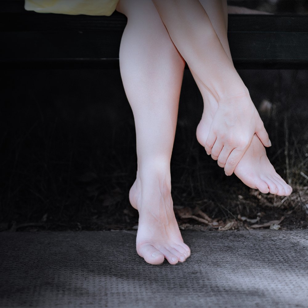
If you are reading this article there is a good chance you have been diagnosed with a stress fracture/bone stress injury (BSI) or are wondering if your symptoms might indicate one. When we hear the word fracture most of us think about a traditional bone break in which a large force applied suddenly to a bone causes it to break. These types of fractures typically result from an accident like a fall or a car crash. Bone stress injuries, however, are quite different in terms of the risk factors, mechanism of injury and treatment approach. Let’s begin by devling a bit further into the anatomy and physiology of a bone stress injury and what symptoms might indicate you have a stress fracture.
INTRO TO BONE STRESS INJURIES
The term bone stress injury is often used when discussing stress fractures. A fracture as it is known in the traditional sense only occurs in later stages of a bone stress injury. Unlike a typical fracture which is usually the result of a sudden, large force, BSIs occur in response to repetitive submaximal loading without sufficient recovery time. This repetitive stress exceeds the bone’s elastic resistance causing structural fatigue and eventually what we know as a stress fracture. There are typically two scenarios that result in a bone stress injury. The first is abnormal stress upon a normal bony structure and the second is normal stress on an abnormal bony structure. In either case, a bone stress injury may occur.
BSIs are most often seen in weight bearing bones. Athletes and military recruits are two of the populations most susceptible to BSIs because of the high volume and repetitive nature of their activities. Running and gymnastics are two sports with higher rates of BSI among athletes. BSIs can be stratified into high and low risk categories based on the likelihood that the fracture will progress to a displaced fracture or non-union fracture and require surgery:
High Risk Stress Fracture Sites
Calcaneus (heel bone)
Fifth metatarsal
Sesamoid bones
Talus
Anterior tibia
Medial malleolus (inside ankle bone)
Neck of the femur
Head of the femur
Patella (knee cap)
Pars articularis of the lumbar spine
Tarsal navicular
Low Risk Stress Fracture Sites
Pubic ramus (lower part of pelvis)
Sacrum
Ribs
Humerus
Posteromedial tibia
Fibula
2nd-4th metatarsal shafts
HOW ARE BONE STRESS INJURIES DIAGNOSED?
If you or your healthcare provider suspects you have a BSI it is important to get imaging to determine the grade or severity of the BSI as this will determine the appropriate treatment plan. While X-rays are usually an excellent choice to diagnose a frank bone fracture, X-rays will typically remain normal in appearance the first few weeks after symptom onset with a BSI. MRI on the other hand is the most sensitive imaging modality and can detect even low grade injuries early. BSIs are staged based on their appearance on MRI (T1 and T2 are different MRI imaging techniques):
Grade 1: Periosteal edema only (periosteum is the outer surface of the bone)
Grade 2: Bone marrow edema (only on T2 weighted sequences)
Grade 3: Bone marrow edema (on T1 and T2 weighted sequences)
Grade 4: (4a) Multiple discrete areas of intracortical signal changes; (4b) Linear areas of intracortical signal change correlating with a frank stress fracture
If a reaction is seen in the outer part of the bone on x-ray it usually correlates with a grade 3 BSI while a fracture line correlates with a grade 4 BSI.
WHAT ARE SYMPTOMS OF A BONE STRESS INJURY
While the symptoms of a BSI may vary slightly based on location, they tend to follow a specific pattern. Typically BSI begins with a complaint of insidious onset of pain that is worse with activity and lessens with rest. If activity continues and the BSI progresses, pain may be felt even at rest. If the area of injury can be palpated by hand, tenderness with palpation of the bone may be experienced over that area. Soft tissue swelling may also be present over the area in the earlier stages. With a stress fracture involving the hip or pelvis, pain may be experienced in the groin and worsened when the leg is rotated inward.
“Shin splints” also known as medial tibial stress syndrome is an indicator of an early stress reaction and if it progresses can be hard to distinguish from a stress fracture.
A “hop test” is commonly used as a tool to identify a BSI, especially in the tibia, as hopping repeatedly on one leg will cause pain caused by BSI but not if it is shin splints. Similarly a “fulcrum test” can be used to identify a femoral shaft stress fracture. Because it may be difficult for you to differentiate a BSI from a soft tissue injury, it is important to seek the advice of a qualified healthcare provider.
WHAT ARE THE RISK FACTORS FOR DEVELOPING A BONE STRESS INJURY?
Risk factors for a BSI include both intrinsic and extrinsic elements that may interact differently within each person during different times in their lives and training. Identifying which risk factors are modifiable versus unmodifiable provides a target to minimize the risk of re-injury in the future. Here are some of the significant risk factors for BSI:
Intrinsic risk factors
Female sex
Older age
Skeletal structure variations
History of prior bone injury
Inadequate muscle strength, flexibility and endurance
Low energy availability (underfueling)
Menstrual cycle dysfunction
Low BMI
Low bone mineral density
Disordered eating
Low Vitamin D
Low calcium stores
Extrinsic risk factors
Inadequate recovery between training sessions
Inadequate sleep duration and quality
Inadequate post-exercise nutrition
Increasing training loads too quickly
Changing training terrain suddenly
HOW CAN PHYSICAL THERAPY HELP?
Physical therapists play an important role in the prevention and recovery from a bone stress injury. Treatment of a BSI depends on its stage and the site of injury. Typically modified weight bearing is prescribed to promote remodeling and rebuilding of the affected area followed by a gradual return to activity once deemed safe by your physician and based on repeat imaging and a reduction in symptoms. While you likely have to take some time off from the sport that contributed to the BSI in the first place, physical therapy should be a part of your recovery from the beginning. Here are some ways that physical therapists address BSI:
Screen for risk factors of BSI and provide counseling, or a referral to another professional for counseling, on how to minimize and modify these risk factors
Prescribe an exercise program for uninjured areas of the body to minimize loss of strength and fitness during recovery time
Create a progressive treatment plan for the injured body part to help it return to full strength and flexibility in preparation for return to sport and activity
Guide you through a return to sport program which includes careful monitoring of symptom response
Address other contributing biomechanical and performance factors that can reduce your risk for re-injury or other injuries in the future
If you are currently recovering from a stress fracture, now is the time to start physical therapy. If you are injured and unsure if you have a stress fracture, our physical therapists can perform an examination to help determine if a referral to an orthopedic or sports medicine physician is needed and then help support you through the recovery process. We are excited to support you in your recovery and return to the sports you love while helping you reduce the likelihood of re-injury in the future.
Click here for more information about Physical Therapy for Bone Stress Fractures
About EvolveNY-
Brooklyn's Premier Holistic Physical Therapy Clinics- There’s physical therapy, there’s training, and then there’s EVOLVE. We use the science of biomechanics merged with fitness to help our patients get better and stay better!
First we evaluate, then we heal, then we strengthen our clients so they can reach their goals, feel better, and live happier lives. We do so by utilizing a range of core techniques and specialized treatments to reduce pain, improve mobility, enhance physical strength and deal with the underlying issues, not just the pain itself.
Multiple Brooklyn Locations!

Physical Therapy for Herniated and Bulging Discs
Intervertebral Disc Herniations 101
Physical Therapy for Herniated and Bulging Discs- The nomenclature around disorders of the intervertebral disc has evolved over time. In the medical field we most often refer to discs as “herniated” or occasionally, “bulging.” Still, you may hear someone say they have a “slipped disc” in a more colloquial conversation. While advanced imaging today makes us pretty certain that discs are not “slipping” in and out of place, disc herniations or bulging discs are certainly a very real cause of neck and back pain treated in a physical therapy clinic.
Get Help With Your Herniated Disc

Intervertebral Disc Herniations 101
The nomenclature around disorders of the intervertebral disc has evolved over time. In the medical field we most often refer to discs as “herniated” or occasionally, “bulging.” Still, you may hear someone say they have a “slipped disc” in a more colloquial conversation. While advanced imaging today makes us pretty certain that discs are not “slipping” in and out of place, disc herniations or bulging discs are certainly a very real cause of neck and back pain treated in a physical therapy clinic.
Symptoms of a herniated disc can include localized pain in the spine, radiating pain into the arms or legs, numbness or tingling in the extremities, pain in the area with coughing or sneezing, weakness in the arms or legs, pain with walking, difficulty sitting for long periods, difficulty standing up straight, pain with bending the spine and bowel/bladder function changes. While the pain and discomfort of a herniated intervertebral disc can be disconcerting, in most cases conservative treatment such as physical therapy can help improve your symptoms.
WHAT IS HAPPENING WHEN A DISC HERNIATES?
Intervertebral discs have an important role to play in the structure of the spine. An intervertebral disc sits between each of the vertebral bodies in the neck, mid back and lower back. They act as a cushion, spacer and shock absorber. The height of the intervertebral disc helps create space for the spinal nerves to pass unencumbered from the spinal cord to the skin, muscles, soft tissue and organs throughout the body. When the height or structure of the disc is altered it can place unwanted pressure on the spinal nerves causing alterations in sensation and muscle function.
If you were to extract an intervertebral disc and examine it you will see that it is fairly oval-shaped and made up of two distinct materials. In the center is the gelatinous nucleus pulposus. Surrounding this area is the strong, fibrous outer layer called the annulus fibrosus. The nucleus helps with shock absorption and cushioning while the annulus helps maintain the shape of the disc and resist the stress and strain applied to it through spinal movement.
In the case of a disc herniation, a tear in the fibrous outer layer allows the gelatinous center of the disc to extrude or protrude outside the confines of the disc. This is what is called a herniated or bulging disc because when seen on imaging, a bulge of nucleus pulposus is seen on the outside of the disc. In most cases disc herniations are a result of natural weakening of the disc as we age but sometimes it can be caused by a single episode of high stress across the back such as picking up something that is too heavy.
Central disc herniations occur when the disc herniates medially toward the spinal cord. This may result in bilateral symptoms depending on the size and location of the bulged disc. Lateral disc herniations can impinge on the spinal nerves as they exit the foramina and will typically cause symptoms localized to one side of the body along the distribution of the affected nerve. The location of symptoms can help your PT or physician determine which intervertebral disc may be involved. While technically a disc herniation can occur anywhere in the spine, they are most common in the cervical spine (neck) and lumbar spine (low back).
Pressure on the spinal nerves or spinal cord caused by the bulging disc material is the cause of the signs and symptoms we associate with a herniated disc. If a herniation occurs in the cervical spine then symptoms are usually experienced in the neck, arms or hands. Alternatively, when a lumbar disc herniates the symptoms may be felt in the lower back, buttock, hips, thighs, lower leg or foot. In severe cases, a large central disc herniation can injure the spinal cord which can lead to more widespread and severe symptoms.
If you are experiencing severe muscle weakness or pain that has not responded to conservative treatment, a surgical consult may be recommended. If you are experiencing sudden loss of bowel or bladder control, immediate surgery is often recommended to decompress the involved nerves.
PHYSICAL THERAPY IS AN EXCELLENT CHOICE TO TREAT A HERNIATED DISC
In all but the most extreme cases physical therapy is a more optimal treatment choice than surgery or opioid pain medication. While initially the pain or discomfort you are experiencing may make you feel like surgery or opioid pain medication are the best choice, a conservative approach like physical therapy can not only provide you with symptom relief but help you avoid the long recovery of a surgery and the potential side effects of pain medication. In most cases an intervertebral disc herniation will spontaneously improve over a period of months but physical therapy can help hasten symptom relief.
In the first couple of days after an acute disc herniation your physical therapist can advise you on strategies to help minimize pain and stiffness. Applying ice to the affected area for 15-20 minutes every couple of hours and making sure you continue to get up and walk throughout the day can help with these symptoms. Similarly, they may advise you on how to modify your sleeping position to find the most comfort or on using a firm chair versus a soft chair. Additionally, while they may recommend avoiding pain-provoking activities at first, it is also important to avoid bed rest because this can actually worsen the pain and stiffness you feel when you decide to get up and get moving again.
After the first couple of days your PT can guide you through an individualized physical therapy program aimed at restoring pain-free motion. They may perform gentle hands-on techniques to restore joint and soft tissue mobility and reduce pain early on. You will learn exercises to help centralize your symptoms so that they no longer radiate into your arms and legs. An individualized strength and flexibility program can help restore strength in any weakened muscles and promote good strength and control around the spine itself. If there are postural habits that may have contributed to the injury you will be instructed in postural exercises as well as training on optimal biomechanics for performing both everyday movements and ones that may place you at higher risk for injury like picking up heavy items.
Treatment for a herniated disc can begin right away. If you are experiencing symptoms of a herniated disc our team of physical therapists is ready to help. Get a jump start on your recovery and the confidence of knowing how to reduce the likelihood of this injury happening again in the future.
Click here for more information about physical therapy for herniated discs
About EvolveNY-
Brooklyn's Premier Holistic Physical Therapy Clinics- There’s physical therapy, there’s training, and then there’s EVOLVE. We use the science of biomechanics merged with fitness to help our patients get better and stay better!
First we evaluate, then we heal, then we strengthen our clients so they can reach their goals, feel better, and live happier lives. We do so by utilizing a range of core techniques and specialized treatments to reduce pain, improve mobility, enhance physical strength and deal with the underlying issues, not just the pain itself.
Multiple Brooklyn Locations!

Physical Therapy for Spinal Stenosis
Seeking Solutions for Spinal Stenosis? Most people will experience back pain at some point in their lives. In most cases, back pain is self-limiting and resolves on its own. Spinal stenosis is one cause of back pain that can lead to persistent or recurring symptoms. In this case, it is a good idea to seek the help of an expert. Research shows conservative care such as physical therapy is an excellent choice to address back pain and spinal stenosis is one of the many causes of back pain addressed at our clinic.
Physical Therapy for Spinal Stenosis
Could Your Pain Be Spinal Stenosis?
Most people will experience back pain at some point in their lives. In most cases, back pain is self-limiting and resolves on its own. Spinal stenosis is one cause of back pain that can lead to persistent or recurring symptoms. In this case, it is a good idea to seek the help of an expert. Research shows conservative care such as physical therapy is an excellent choice to address back pain and spinal stenosis is one of the many causes of back pain addressed at our clinic. In this article we will delve deeper into spinal stenosis, how it is diagnosed and how physical therapy can address your symptoms in both the short and longer term.
WHAT IS SPINAL STENOSIS AND HOW DOES IT FEEL?
Spinal stenosis refers to a narrowing of the spaces created by the vertebrae in the spinal column causing pressure on the delicate nerves. It typically occurs in the neck (cervical spine) and lower back (lumbar spine). When narrowing occurs in the central spinal canal that houses the spinal cord it is called central stenosis. This can put pressure on the spinal cord itself. When narrowing occurs on the lateral portion of the vertebrae it is called lateral stenosis and may put pressure on the spinal nerves that exit from the spinal canal and travel out to the skin, bones, joints, muscles and organs. This pressure causes the classic symptoms of spinal stenosis including pain in the neck and/or back pain, numbness, tingling or weakness in the arms, shoulders, legs or trunk possibly accompanied by intermittent bowel and bladder problems.
In many cases stenosis develops as a natural result of aging. Loss of intervertebral disc height, arthritis and the development of bone spurs can all cause spinal stenosis. In some cases stenosis may be the result of thickening of the spinal ligament or medical conditions like Paget disease or a spinal tumor.
Because of the narrowing, many people with spinal stenosis find they are most comfortable when their spine is flexed such as when sitting. Alternatively, they find it difficult to walk or stand for long periods of time. At the grocery store, for example, many people with spinal stenosis will find themselves leaning over their shopping cart as this relieves some of the nerve compression created by the stenosis and allows them to shop longer. This is called the “shopping cart sign”.
HOW IS SPINAL STENOSIS DIAGNOSED?
Many times patients come to physical therapy already diagnosed with spinal stenosis. In some cases, however, a physical therapist may be the first one to suspect this condition. One symptom in particular may lead PT’s to suspect spinal stenosis. If an individual complains of pain, tingling or cramping, weakness or heaviness in the hips, legs or buttocks with activity, spinal stenosis should be on the differential. Two different conditions may be the culprit here.
One condition called vascular claudication is a symptom of peripheral artery disease. Peripheral artery disease causes narrowing of the vessels that carry blood and oxygen to working muscles. Decreased blood flow caused by this narrowing can lead to the symptoms listed above. Alternatively, neurogenic claudication is the result of compression of the spinal nerves caused by spinal stenosis. Because these conditions can be difficult to differentiate by symptom report, a test called the Bicycle Test of Van Gelderen was developed to provide some further insight into the underlying cause. During this test an individual rides a bicycle in an upright position and a forward leaning position. If the individual experiences symptoms of claudication in both positions then a vascular origin is suspected. If symptoms are experienced while upright and relieved while leaning forward then spinal stenosis is suspected as the forward leaning position helps relieve compression and irritation of the spinal nerves.
Imaging of the spine is the most definitive way to diagnose spinal stenosis. X-rays, MRIs and CT scans can all be used to evaluate the spine. By looking at the bony and soft tissue structures of the spine doctors and therapists can evaluate the alignment of the spine and look for areas where the spinal canal or the foramina (holes created by the vertebrae through which spinal nerves exit the spinal canal) are narrowed. In some cases, narrowing of the spinal canal or foramina found on imaging may correlate directly with symptoms like pain with prolonged standing. Other times, the findings are incidental and despite the narrowing, do not present any problems for the patient. This is important to remember when looking at imaging studies. Not every so-called abnormality found on imaging results in symptoms so it is important to take into account a full patient history and exam to determine if other factors not shown on imaging should be addressed instead or additionally.
IS PHYSICAL THERAPY USEFUL IN TREATING SPINAL STENOSIS?
The short answer is yes! While physical therapy cannot reverse arthritis or increase the height of your intervertebral discs, research shows that conservative treatment like physical therapy can be useful in managing the symptoms of spinal stenosis. The goal of physical therapy for stenosis is to increase your tolerance for functional activities like walking, climbing stairs, standing and so forth. While each patient will receive an individualized physical rehabilitation program depending on their needs and goals, there are some common interventions used in the treatment of stenosis.
Your therapist may apply some gentle hands-on manual therapy to the muscles, joints and soft tissue to optimize alignment and mobility while also providing pain relief. They will create a program of flexibility and strengthening exercises to help provide good alignment and support to the spine. You will be educated on strategies for modifying your posture or activities to reduce discomfort and possibly on how to integrate an assistive device into your routine to improve your ability to stand and walk.
If you have been diagnosed with spinal stenosis and symptoms are limiting your ability to perform daily activities it is time to take action. Call and schedule an initial evaluation with Evolve Physical Therapy today and let our team of expert physical therapists help you achieve symptom reduction and improve your daily function.
Click here to find out more information about physical therapy for spinal stenosis
About EvolveNY-
Brooklyn's Premier Holistic Physical Therapy Clinics- There’s physical therapy, there’s training, and then there’s EVOLVE. We use the science of biomechanics merged with fitness to help our patients get better and stay better!
First we evaluate, then we heal, then we strengthen our clients so they can reach their goals, feel better, and live happier lives. We do so by utilizing a range of core techniques and specialized treatments to reduce pain, improve mobility, enhance physical strength and deal with the underlying issues, not just the pain itself.
Multiple Brooklyn Locations!
https://EvolveNY.com

Physical Therapy for Sciatica
Physical Therapy for Sciatica- There is no mistaking the pain of sciatica. Whether it’s shooting down the back of your leg or creeping up on you during a long car ride, you’ll quickly find yourself searching for a position or stretch to provide relief. As many as 40% of people will experience sciatica at some point in their lives which means at any given time there are many people looking not only for immediate relief but for long-term solutions. In this article we will discuss the common presentation of sciatica, its underlying causes and how physical therapy can treat this condition.
Is Sciatica a Pain in Your Butt?

There is no mistaking the pain of sciatica. Whether it’s shooting down the back of your leg or creeping up on you during a long car ride, you’ll quickly find yourself searching for a position or stretch to provide relief. As many as 40% of people will experience sciatica at some point in their lives which means at any given time there are many people looking not only for immediate relief but for long-term solutions. In this article we will discuss the common presentation of sciatica, its underlying causes and how physical therapy can treat this condition.
IS MY PAIN REALLY SCIATICA?
Sciatica refers to symptoms caused by irritation or compression of the sciatic nerve. When this nerve is irritated, symptoms will present themselves in the back of the thigh, leg and foot where the sciatic nerve and its branches provide motor and sensory input to the leg. A lot of people are familiar with the deep ache or shooting pain that can occur down the back of the thigh but these are not the only symptoms of sciatica. If you are experiencing any of the following symptoms, there is a good chance that sciatica may be the culprit:
Pain and/or pressure in the back, hip, leg(s), foot/feet
Throbbing, aching, shooting, sharp, dull, or burning pain in the buttock, back of the thigh, lower leg or foot
Numbness or tingling in the back, hip, leg, or foot
Weakness in the leg(s)
Inability to bend or rotate the back
Increased pain when coughing, sneezing, reaching, or sitting
Inability to stand up straight
Difficulty getting up from a chair due to pain or weakness
Inability to remain in one position for a long period of time, such as sitting or standing, due to pain
Pain that is worse in the morning
Limping when walking
The sciatic nerve is not the only nerve that can refer pain, numbness, tingling or weakness into the leg and a physical therapist can help localize the involved spinal nerves and segments based on the location of your symptoms. Identifying the origin of your symptoms will ensure that the treatment or exercises you do to address the problem are appropriate.
LET’S TAKE A LOOK AT SOME OF THE UNDERLYING CAUSES OF SCIATICA
Getting to the root of your symptoms is one important way that physical therapists can help facilitate long term resolution of your symptoms. A one-size-fits-all approach may provide some relief but is unlikely to take care of your problem entirely. In order to know how to treat these conditions we need to know what causes them. Here we will discuss some of the different causes of sciatica.
Let’s start with a review of basic spine anatomy. The sciatic nerve exits the spine through holes, or foramina, in the lumbar vertebrae of the low back. The sciatic nerve is a bundle of nerve roots from the last two lumbar segments (L4-L5) and upper sacral nerves (S1-S3). It travels down the back of the thigh splitting into smaller nerves that continue on down the lower leg and into the foot. It provides sensation and motor information to these areas and to the structures involved in bowel and bladder function. Sudden loss of bowel or bladder control can be a symptom of severe sacral nerve compression and is a medical emergency.
Sciatica, also called lumbar radiculopathy or “pinched nerve” can have many causes. A bulging or herniated disc, for example, can put pressure on the sciatic nerve roots as they exit the spinal column causing radiating symptoms into the legs. Similarly, bone spurs or arthritis that narrow the foraminal exit for these nerves may cause some irritation especially with certain movements or prolonged positioning. Acute injuries like a muscle strain can cause localized inflammation of tissue over the nerves causing compression and irritation. Finally poor posture or repeated movements may be a contributing factor to sciatica if biomechanics, strength and flexibility aren’t optimal.
WHY SHOULD I CHOOSE PHYSICAL THERAPY TO HELP ME WITH THIS CONDITION?
As you can see, there are many potential causes of sciatic pain. This is why it is so important to work with a skilled physical therapist if you have been unable to manage or fully eliminate these symptoms on your own. Through a careful interview and physical exam you will receive an individualized physical therapy treatment plan that will take into account your body’s unique presentation as well as the specific activities and exercises your daily life requires.
So, while no two treatment plans for sciatica will be exactly the same, these are some of the goals that will likely be a part of your plan of care:
Improve spinal mobility: Ensuring that proper flexibility and mobility exists in the entire spine is important to help balance stress and strain through the lower back. This can help reduce irritation or compression of the sciatic nerve roots.
Address mobility limitations in other areas of the body: While the pain may be originating in your spine, we can’t forget that the body is connected from head to toe. If you are lacking mobility or flexibility in the shoulders, hips, or ankles, for example, you may be overcompensating with your spine. Failure to address these areas may mean you continue to have extra stress through the lower lumbar spine.
Optimize spinal control: The spine was meant to move, but if it is not moved in a controlled manner, conditions like sciatica can develop. Learning to perform movements with the arms and legs while controlling the movement of the spine can not only improve your overall performance but also reduce symptoms of back pain or radiating pain.
Build strength and endurance to support the spine: Strength training of the abdominals, pelvic floor, hips and other areas is often an important tool in treating sciatica.
Learn to move better: If repetitive movements or poor posture are contributing to your sciatica symptoms then learning how to optimize your movements is an important step in facilitating long term results and minimizing the risk of symptom recurrence.
So, as you can see, a comprehensive treatment plan for sciatica is more than just performing a few stretches. This is why physical therapy is an excellent choice for treating sciatica. Let our team of therapists take the reins and help you achieve long-lasting relief of your sciatica symptoms.
Click here for more information about physical therapy for sciatica.
About EvolveNY-
Brooklyn's Premier Holistic Physical Therapy Clinics- There’s physical therapy, there’s training, and then there’s EVOLVE. We use the science of biomechanics merged with fitness to help our patients get better and stay better.
First we evaluate, then we heal, then we strengthen our clients so they can reach their goals, feel better, and live happier lives. We do so by utilizing a range of core techniques and specialized treatments to reduce pain, improve mobility, enhance physical strength and deal with the underlying issues, not just the pain itself.
Multiple Brooklyn Locations!
